Urinary Bladder is one of the organs of the human body located in the anterior part of the pelvis, which is also a muscular reservoir of urine. Urinary bladder is generally included in clinical circumstances like retention of urine, cystitis (inflammation of the urinary bladder), calculus, disorder of micturition, and cancer. The clinicians find the anatomy of urinary bladder, of enormous relevance.
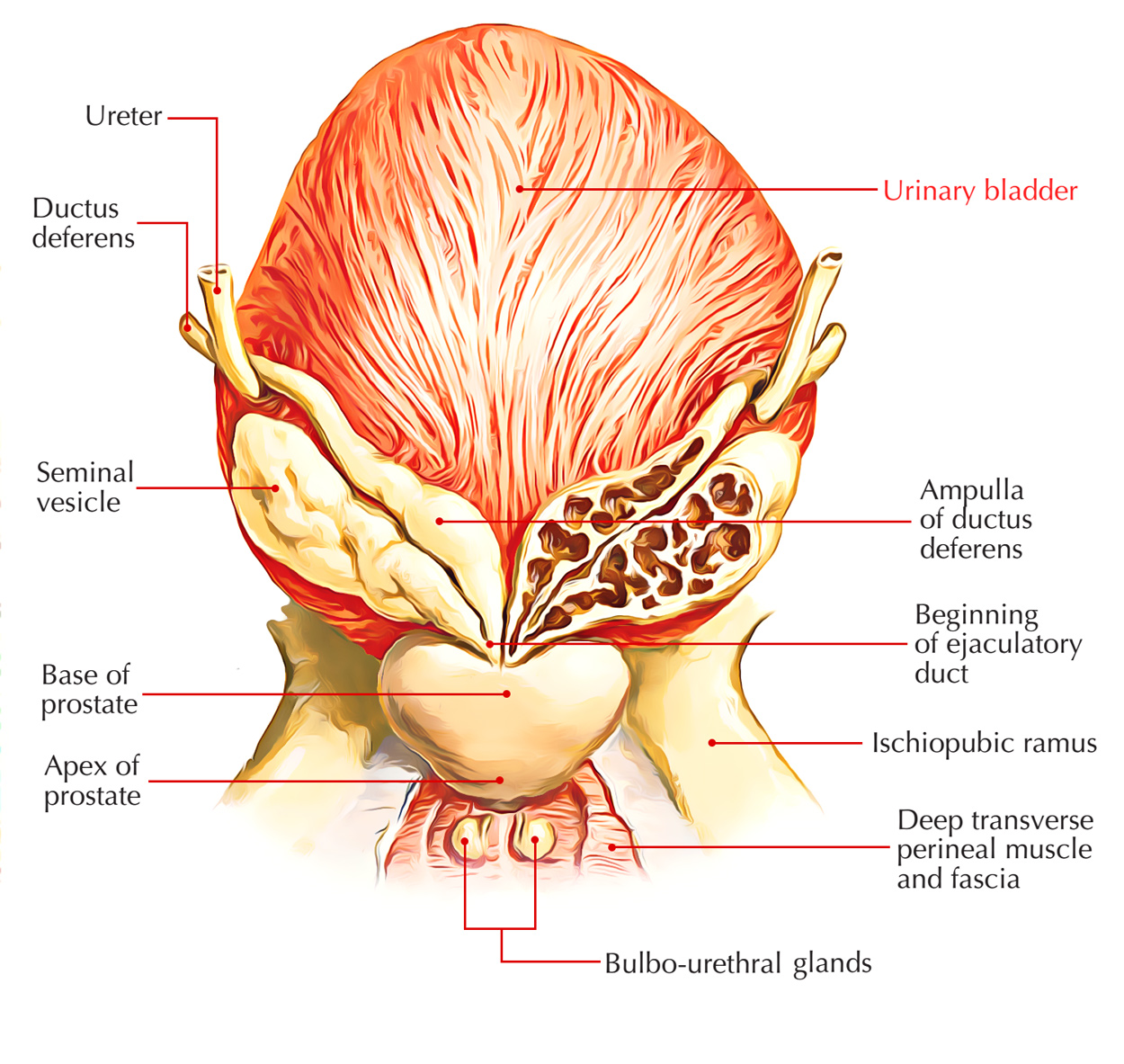
Urinary Bladder
Location
The location of urinary bladder in males is in front of the rectum and in females, in front of the uterus. Urinary bladder is situated in the anterior part of the lesser pelvis instantly behind the pubic symphysis. There is a change in the location of the urinary bladder according to the age of the person and the amount of urine, the urinary bladder is carrying.
When the urinary bladder is empty it is located wholly inside the lesser pelvis however when it becomes distended with urine, it grows upward and forward into the abdominal cavity.
In kids, the bladder is an abdominopelvic organ even when it’s empty as the pelvic cavity is small and the neck of bladder is located at the level of the upper border of pubic symphysis. Urinary bladder starts to go into the enlarging pelvis in the age of 6 years but doesn’t become a pelvic organ thoroughly until after puberty.
Size and Shape
The size and shape of the urinary bladder change based on the amount of pee it includes. Urinary bladder is tetrahedral in shape when empty and oval in shape when distended.
Capacity
Normally in adult male the capacity changes from 120 to 320 ml. The mean capacity is all about 220 ml.
- An amount of urine beyond 220 ml causes a desire to micturate but the bladder is normally emptied at about 250-300 ml.
- The filling of pee up to 500 ml could be endured but beyond this, it causes pain because of tension of its wall. On collection of pee about 800 ml, the micturition is beyond one’s voluntary control.
Functions
There are 2 functions of the urinary bladder:
Temporary store of urine: The walls of the urinary bladder are expandable, with folded internal lining which allows the urinary bladder to hold upto 600ml.
Assist In displacement of urine: At the time of voiding, the musculature of the urinary bladder contracts, and the sphincters relax.
External Features and Relations
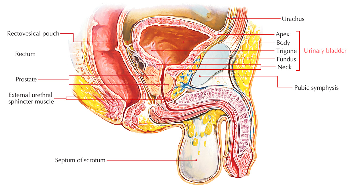
Urinary Bladder: Features
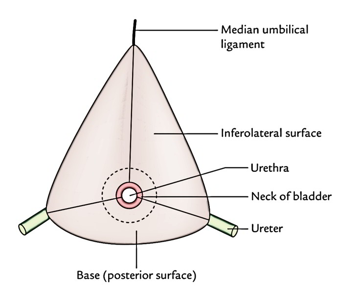
An empty and caught bladder as observed in a cadaver istetrahedral in shape and presents the following external features:
- Apex.
- Base.
- Neck.
- 3 surfaces (superior and 2 inferolateral surfaces).
- 4 edges (anterior, posterior and 2 lateral).
Urinary Bladder: Features
Apex
It gives connection to the median umbilical ligament and is located posterior to the upper margin of the pubic symphysis.
The median umbilical ligament is the fibrous remnant of the intra-abdominal part of the allantois (urachus).
Base (Posterior Surface/Fundus)
- The urinary bladder is triangular in shape and directed posteroinferiorly toward the rectum.
- Its superolateral angles are joined by the ureters while its inferior angle provides rise to the urethra.
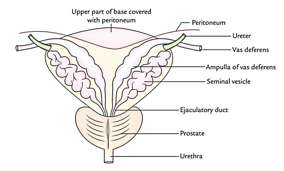
In the male, its relationships are:
- Upper part is divided from rectum by the rectovesical pouch consisting of coils of the small intestine.
- Lower part is divided from rectum by the terminal parts of vasa deferentia and seminal vesicles.
- The vasa deferentia is located medial to the seminal vesicles.
- The triangular area between the vasa deferentia is divided from the rectum by rectovesical fascia (of Denonvilliers).
Urinary Bladder: Base
In the female, it’s split from the cervix of uterus and by the vesicouterine pouch.
Neck
It’s the lowest and most fixed part of the bladder. It’s situated where the inferolateral and the posterior surfaces of the bladder meet. It’s pierced by the urethra. It is located about 3-4 cm behind the lower part of pubic symphysis. Its Relations are:
- In the male, it rests on the upper surface of the prostate where the smooth muscle fibres of the bladder wall are constant with those of the prostate.
- In the female, it’s related to the urogenital diaphragm.
Superior Surface
It’s triangular in shape and jump on every side by the lateral edges which stretch from ureteric orifices posterolaterally to the apex anteriorly and posteriorly by the posterior border which joins the ureteric orifices.
In the male, it’s fully covered by the peritoneum which divides it from:
- coils of the ileum, and/or.
- sigmoid colon.
Along its lateral edges, the peritoneum is reflected on to the pelvic walls.
In the female, it’s covered by the peritoneum with the exception of a small area near the posterior border, that is related to the supravaginal part of the uterine cervix. Here the peritoneum is reflected on to the uterine isthmus creating vesicouterine pouch.
Inferolateral Surfaces
Every inferolateral surface slopes downward, forward, and medially to meet its fellow of the reverse anteriorly in the midline.
These surfaces are divided from every other, anteriorly by the anterior border, and from the superior surface by the lateral edges.
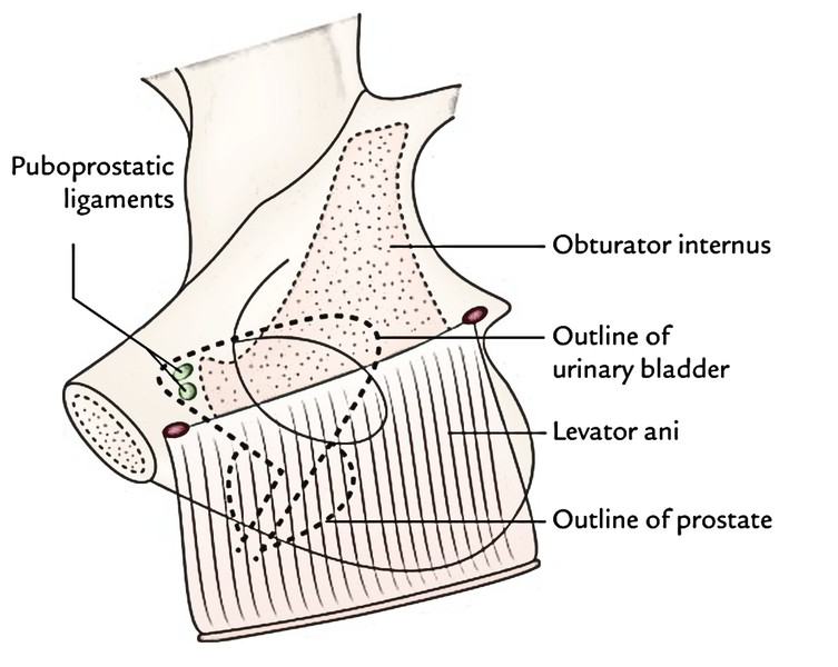
The inferolateral surfaces are devoid of peritoneum and in both male and female are associated:
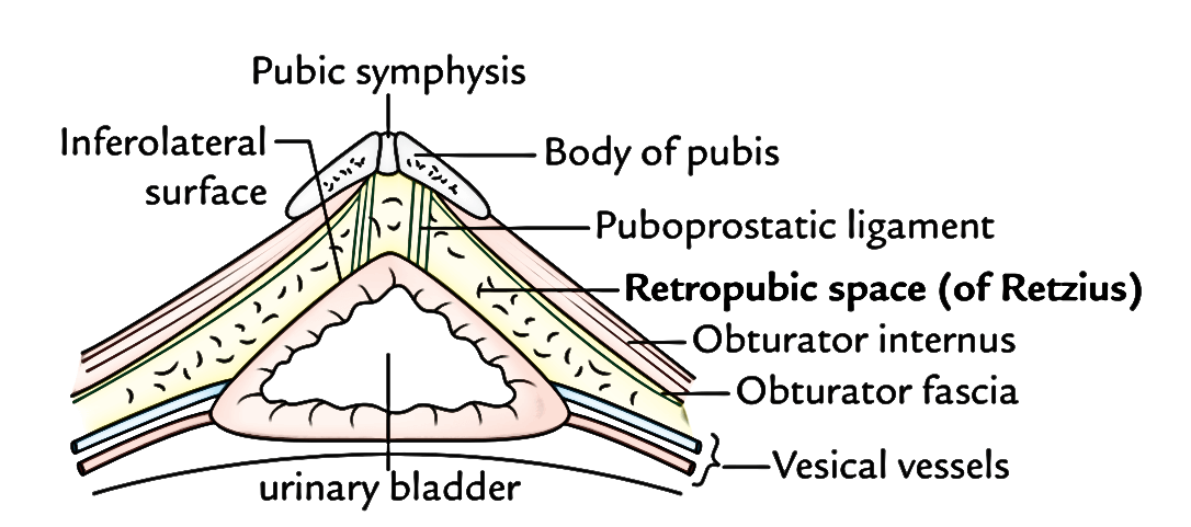
Urinary Bladder: Inferolateral Surface in Females
In front to.
- retropubic space,.
- pubic symphysis, and.
- puboprostatic ligaments.
Urinary Bladder: Inferolateral Surface in Males
Behind to.
- obturator internus muscle above, and.
- levator ani muscle below.
Retropubic space (cavern of Retzius): It’s a perivesical space bounded anteriorly by the posterior aspect of pubic symphysis, and adjoining posterior wall of rectus sheath; posteriorly by inferolateral surfaces of the urinary bladder, superiorly by reflection of peritoneum from the superior surface of urinary bladder to the posterior aspect of the anterior abdominal wall up to the umbilicus, and inferiorly by puboprostatic/pubovesical ligaments.
Relationships of the Urinary Bladder
| Parts | Relations |
|---|---|
| Base | 1. Rectovesical pouch in the male 2. Vesico uterine pouch in the female 3. Vasa deferentia and seminal vesicles (separated from the rectum by fascia of Denonvilliers) |
| Superior surface | 1. Peritoneal cavity containing loops of ileum 2. Coils of ileum 3. Sigmoid colon 3. Uterine cervix (in female) |
| Anterior surface (inferolateral surfaces) | 1. Retropubic space 2. Puboprostatic ligaments 3. Obturator internus and levator ani muscles |
| Apex | Median umbilical ligament |
| Neck | 1. Prostate gland (in male) 2. Urogenital diaphragm (in female) |
Clinical Significance
Suprapubic aspiration of the urinary bladder: When the urinary bladder is distended it peels off the peritoneum from the anterior abdominal wall and comes in its direct contact. Now it can be aspirated suprapubically without any damage to the peritoneum. In suprapubic cystostomy, the exact same state can be gotten by man-made distension of the urinary bladder.
Supports of the Urinary Bladder (Fixation of the Urinary Bladder)
The urinary bladder is anchored securely at its neck, where it’s fixed by its continuity together with the prostate and urethra. The fixation of the bladder is also helped by the various ligaments of the urinary bladder.
Ligaments
The ligaments of the bladder are of 2 types – true and fictitious.
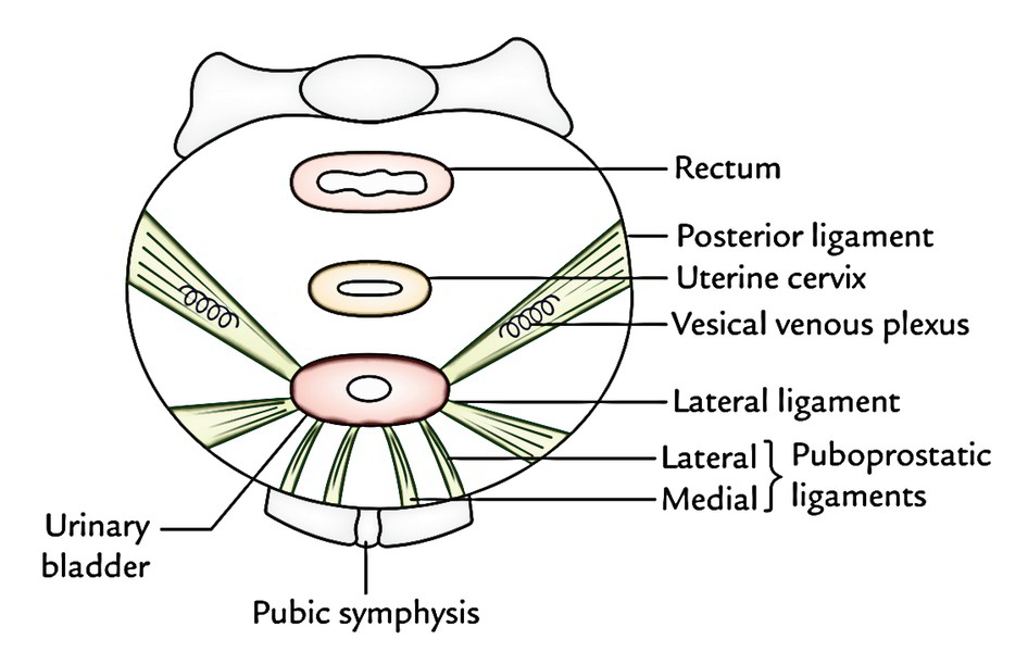
Urinary Bladder: Ligaments
True Ligaments
All these are created by the condensation of pelvic fascia around the neck and the base of the bladder and have a supporting function for the bladder.
- Lateral ligaments (2 in number, left and right): They stretch from the side (inferolateral surface) of the bladder to the tendinous arch of pelvic fascia.
- Puboprostatic ligaments (4 in number, 2 on every side- medial and lateral): They fix the neck of bladder.
- Lateral puboprostatic ligament goes downward and medially from the anterior end of the tendinous arch of pelvic fascia to combine together with the upper part of the prostatic sheath.
- Medial puboprostatic ligament extends downward and backward from the rear of the pubic bone near the pubic symphysis to the prostatic sheath and creates the floor of retropubic space (of Retzius).
Fascial bands quite similar to puboprostatic ligaments in the female are referred to as pubovesical ligaments. They stop around the neck of the urinary bladder.
- Median umbilical ligament is the fibrous remnant of the urachus. It goes from the apex of the bladder to the umbilicus. It keeps the bladder in position anteriorly and superiorly.
- Posterior ligament (2 in number, left and right): They go as a sheet of loose areolar tissue from the side of the base of the bladder to the lateral pelvic wall. They enclose the vesical venous plexus.
Bogus Ligaments
All these are peritoneal folds and don’t have supporting function as performed by true ligaments. They’re 7 in number.
- Anteriorly there are 3 folds:
a) Median umbilical fold, the fold of peritoneum over the median umbilical ligament.
b) 2 medial umbilical folds, the folds of peritoneum over the obliterated umbilical arteries.
- Laterally a pair of bogus lateral ligaments is composed by the reflection of the peritoneum from the bladder to the side wall of the pelvis and creates the floor of paravesical fossae.
- Posteriorly a pair of bogus posterior ligaments is composed. All these are the sacrogenital folds that are the folds of peritoneum stretching from the side of the bladder, posteriorly, on each side of the rectum, to the anterior aspect of the 3rd sacral vertebra.
Microscopic Structure
The bladder wall from inside outward consists of:
- A mucous membrane.
- A muscular layer.
- Adventitia.
Mucous Membrane
It’s light pink in colour and covered using a transitional epithelium. It’s thrown into folds (rugae) when the bladder is empty. The mucosal area covering the internal outermost layer of the base of the bladder is referred to as “trigone”.
Muscular coating makes up the detrusor muscle which is composed of 3 layers of smooth muscle fibres:
- An outer longitudinal layer.
- A middle circular layer.
- An inner longitudinal layer.
There’s profuse intermingling of the muscle fibres of these layers and they can’t be divided into 3 clearly defined layers.
Since the muscle fibres of the bladder wall are primarily concerned with the evacuation of the bladder they’re together termed the “detrusor muscle”.
Adventitia
It’s created from fibroelastic tissue.
Inside of The Bladder
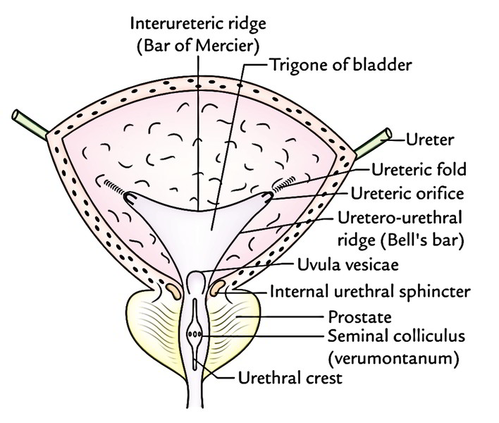
Urinary Bladder: Inside
- In an empty bladder, the greater part of mucosa demonstrates atypical folds (rugae) because it’s loosely connected to the subjacent muscular layer.
- Over a small triangular area, immediately above and behind the internal orifice of the urethra (trigone of the bladder), the mucous membrane is securely bound to the muscular layer and hence is smooth. The limits of trigone are defined superiorly by the openings of the ureters and inferiorly by the urethra.
The trigone of urinary bladder presents the followingfeatures:
- Anteroinferior angle, created by the internal orifice of the urethra.
- 2 posterosuperior angles, created by openings of the ureters.
- Uvula vesicae, a small elevation in the mucous membrane immediately above and behind the internal urethral orifice. It’s generated by the median lobe of prostate.
- Interureteric ridge/crest (bar of Mercier) creates the superior boundary of trigone and attaches both ureteric orifices. It’s created by the continuance into the bladder wall of the inner longitudinal coatings of the ureters.
The lateral ends of the ridge stretch past the openings of the ureter as ureteric folds (created by the ureters as they run obliquely via the bladder wall).
The interureteric ridge (pub of Mercier) acts as a guide to find the orifices of the ureter during cystoscopy.
- 2 uretero-urethral ridges (Bell’s bars) go from the ureteric orifice to the urethral orifice. They’re made by longitudinal fibres of the ureter which goes behind the ureteric orifice down on every side of trigone toward the middle lobe of the prostate.
Arterial Supply
It’s by the following arteries:
- The main arteries supplying blood to the bladder are superior and inferior vesical arteries that are the branches of anterior section of internal iliac arteries.
- The other arteries which make small contribution in supplying the lower part of the bladder are:
- Obturator and inferior gluteal arteries.
- Uterine and vaginal arteries in the female.
Venous Drainage
The veins of the bladder don’t follow the arteries. They create a complex plexus on the inferolateral surfaces near the prostate referred to as vesical venous plexus.
- This plexus enters backwards in the posterior ligaments of the urinary bladder to drain into the internal iliac veins.
- It interacts:
- In the male together with the prostatic venous plexus.
- In the female with the veins at the base of broad ligament.
Lymphatic Drainage
The lymphatics of the bladder drain mainly into the external iliac lymph nodes. Some lymph vessels also drain into the internal iliac lymph nodes consisting of nodes in the obturator fossa.
Nerve Supply
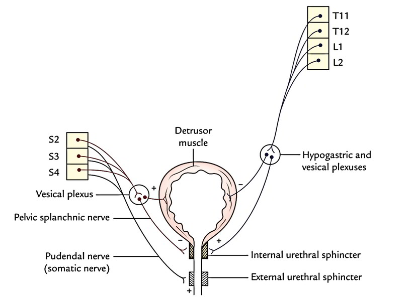
Urinary Bladder: Nerve Supply
Motor Innervation
It’s supplied by the parasympathetic, sympathetic, and somatic fibres.
- Parasympathetic fibres (nervi erigentes) are originated from S2, S3, S4 (spinal micturition center) sections of the spinal cord. They’re motor to the detrusor muscle and inhibitory to the sphincter vesicae (internal urethral sphincter).
- Sympathetic fibres are originated from T11, T12 thoracic and L1, L2 lumbar sections of the spinal cord.
They’re inhibitory to the detrusor and motor to the sphincter vesicae.
- Somatic fibres (pudendal nerve) are originated from S2, S3, S4 spinal sections. They’re motor to the external urethral sphincter.
The sympathetic innervation accounts for the filling of the bladder and parasympathetic innervation for the emptying of the bladder. The somatic innervation is accountable for voluntary constraint of micturition.
Sensory Innervation
Nearly all sensory fibres run along the parasympathetic fibres (pelvic splanchnic nerves/nervi erigentes; S2, S3, S4). Some fibres also run with all the sympathetic fibres. The division of sympathetic fibres (presacral neurectomy will not relieve bladder pain because pain fibres are carried by both sympathetic and parasympathetic fibres.
2 types of fibres are understood:
- Fibres concerned with pain.
- Fibres concerned with conscious knowledge of filling of the bladder.
- The pain fibres run in the anterolateral white Pillars of the spinal cord.
- The fibres concerned with the recognition of filling of the bladder is located in the posterior Pillars of the spinal cord.
Medically, this accounts for the reality that consciousness of the bladder being filled up and want to micturate stay normal after bilateral anterolateral cordotomy for the alleviation of pain.
Clinical Significance
Trabeculated Bladder
It results because of chronic obstruction to the outflow of the pee by enlarged prostate or stricture of the urethra. The bladder becomes distended and its musculature hypertrophies. Its muscular fasciculi boost in size and interface in all the directions providing rise to an open weave look of the bladder wall, called the “trabeculated bladder”.
Suprapubic Cystostomy
It’s an extraperitoneal method to open the cavity of the urinary bladder. It’s done for: Drainage functions, treatment of intravesical circumstances, viz., vesical stones, etc., and removal of the prostate.
The bladder is distended (if not the case of retention of urine) with about 300 ml of fluid. Consequently, the anterior aspect of bladder comes in direct contact together with the anterior abdominal wall. The bladder can be now approached via anterior abdominal wall without entering into the peritoneal cavity.
Neurogenic Bladder
Micturition is basically a reflex action affecting the sensory and motor (sympathetic and parasympathetic) nerve pathways being mediated by the lower micturition center (S2, S3, S4). The voluntary control over this reflex is used by the higher center (cerebral cortex) via upper motor neurons of pyramidal tract.
- Any defect in this nervous mechanics of micturition results in neurogenic bladder.
- The neurogenic bladder lacks the normal nerve charge of micturition.
- The types of neurogenic bladder are:
- Automatic reflex bladder: It results from complete transection of the cord above the lower micturition center (S2, S3, S4) affecting pyramidal tracts (upper motor neurons) medically it presents as:
- The voluntary inhibition and initiation of micturition are lost.
- The bladder empties reflexly every 1 to 4 hours. When the fill reaches a particular level, the detrusor muscle contracts reflexly as in early infancy. This is termed automatic or reflex bladder.
- Sovereign bladder: It Results from the destruction of S2, S3, S4 sections of the spinal cord (lower center of micturition). Medically it presents as:
- 1. The reflex and nervous constraint of micturition is lost. The bladder wall becomes flaccid and capacity of bladder is significantly raised. The end result is constant dribbling and this type of bladder is known as an autonomous bladder.
- 2. The bladder might be emptied by the manual compaction or by the abdominal muscular contraction.
- Gap of sensory side of reflex (example, tabes dorsalis) leads to atonic neurogenic bladder with remaining pee. A large amount of urine accumulates without any reflex contraction.
- In lesions above S2 and below T4 T6, the sympathetic innervation is complete and the patient for that reason becomes conscious when the bladder is full.
Development
The urinary bladder grows from these sources:
- Entire of bladder with the exception of its apex grows from vesicourethral canal (upper part of urogenital sinus).
- Apex of the bladder is originated from the proximal part of the allantoic diverticulum.
- Mucous membrane of trigonum vesicae is originated from the mesoderm of the comprised lower ends of the mesonephric ducts.
- Mucous membrane in the remaining part of the bladder is originated from the endoderm of the vesicourethral part of the cloaca.
- Muscle and serous coat of the bladder are originated from the splanchnic layer of the lateral plate mesoderm.
Clinical Significance
- Congenital anomalies are flawed obliteration of urachus.
- Urachal fistula: The urachus is the abdominal part of allantois going from the apex of the bladder to the umbilicus. It normally obliterates and creates the median umbilical ligament but scarcely stays obvious leading to the urachal fistula, which can lead to discharge of the pee via umbilicus. Medically it presents as discharge of urine from the umbilicus.
- Occasionally the intermediate part of the urachus neglects to obliterate and creates the urachal cyst.
- If distal part of urachus neglects to fibrose, it results in formation of urachal sinus.

 (68 votes, average: 4.57 out of 5)