The eyelids, a.k.a palpebrae, are movable drapes in front of the eyeball. The eye is shielded by them from injury, foreigodies and bright light, keep the cornea moist and clean. Palpebral fissure is the space between both eyelids. The medial angle of the palpebral fissure where two eyelids meet is named medial canthus of the eye and lateral angle of the palpebral fissure where two eyelids meet is referred to as lateral canthus of the eye.
The free margin of every eyelid is split into two parts:
- Sidelong 5/6th ciliary part that is flat and possesses eyelashes and
- Medial 1/6th lacrimal part that is round and doesn’t possess cilia.
Structure
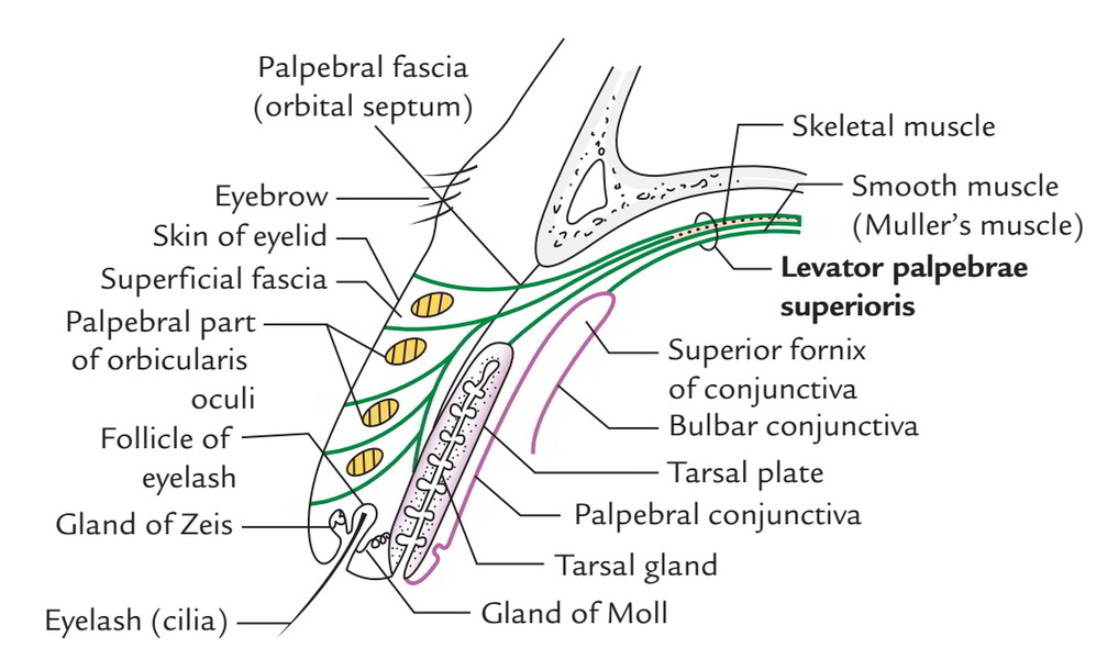
Every eyelid includes 5 layers. From without inwards these are:
- Skin.
- Superficial fascia.
- Orbicularis oculi (palpebral fibres).
- Tarsal plate and palpebral fascia.
- Conjunctiva.
1. Skin: The skin of eyelids is extremely thin and without hair with the exception of at the eyelid margin. At the eyelid margin, it becomes constant with the conjunctiva.
2. Superficial fascia: The superficial fascia of eyelids is thin, free and devoid of fat. It enables the skin to move freely over the eyelid and can become considerably swollen with fluid or blood after injury.
3. Orbicularis oculi: The fibres of palpebral part of orbicularisoculi sweep across the eyelids parallel to the palpebral fissure. A layer of loose areolar tissue is located deep to these fibres and in the upper eyelid it’s constant with the subaponeurotic space of the entire scalp.
4. Tarsal plate and palpebral fascia: The tarsi are two thin plates of conholesed fibrous tissue, which create the skeleton of the eyelids and supply them rigidity. The inferior tarsal plate is a narrow strip connected to the inferior orbital margin by palpebral fascia. The superior tarsal plate is a lot bigger and diamond shaped and can be felt if the upper eyelid is crimped sidelong between finger and thumb.
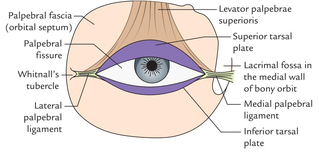
The medial ends of tarsi are connected to lacrimal crest of maxilla in front of lacrimal sac by a strong fibrous band referred to as medial palpebral ligament and lateral ends of tarsi to a tubercle of zygomatic bone (Whitnall’s tubercle) by lateral palpebral ligament.
The large altered sebaceous glands (Meibomian or tarsal glands) are partially embedded on the deeper aspects of the tarsal plates. These glands are ordered within row and their ducts open into the eyelid margin by tiny foramina behind the eyelashes.
The tarsal glands secrete fatty fluid that reduces evaporation of tears and keep them from overflowing onto the cheek.
The ciliary glands are arranged in a number of rows immediately behind the root of eyelashes. Their ducts open on the eyelid margin close to the lashes.
The ciliary glands are of 2 types: (a) glands of Zeis that are changed sebaceous glands and open into the follicles of eyelashes and (b) glands of Moll that are modified sweat glands. They also open into the follicles of the eyelashes.
The palpebral fascia of upper eyelid is connected above to the superior orbital margin and below to the anterior outermost layer of the tarsal plate some space far from its upper border.
The palpebral fascia is the thin fibrous membrane, which attaches the tarsi to the orbital margins and creates the orbital septum with them. Medially it enters posterior to the lacrimal sac and connected to the posterior margin of the lacrimal groove, which lodges the lacrimal sac.
The upper palpebral fascia in the upper eyelid is pierced by:
- Fibres of levator palpebrae superioris,
- Palpebral part of lacrimal gland and
- Nerves and vessels that pass from orbit to the face.
Conjunctiva (palpebral part): It’s a see-through mucus membrane, which lines the inner surface of every eyelid. About 2 millimeters from the border of every eyelid the palpebral conjunctiva presents a groove where foreign bodies often stay.
Arterial Supply
The arterial supply is by medial palpebral branches of ophthalmic artery and lateral palpebral branches of lacrimal artery. These branches create an arterial arch in every eyelid.
Venous Drainage
The venous blood from eyelids is emptied into ophthalmic and facial veins.
Lymphatic Drainage
The lymph from medial halves of eyelid is emptied into submandibular lymph nodes and from lateral halves into the preauricular lymph nodes.
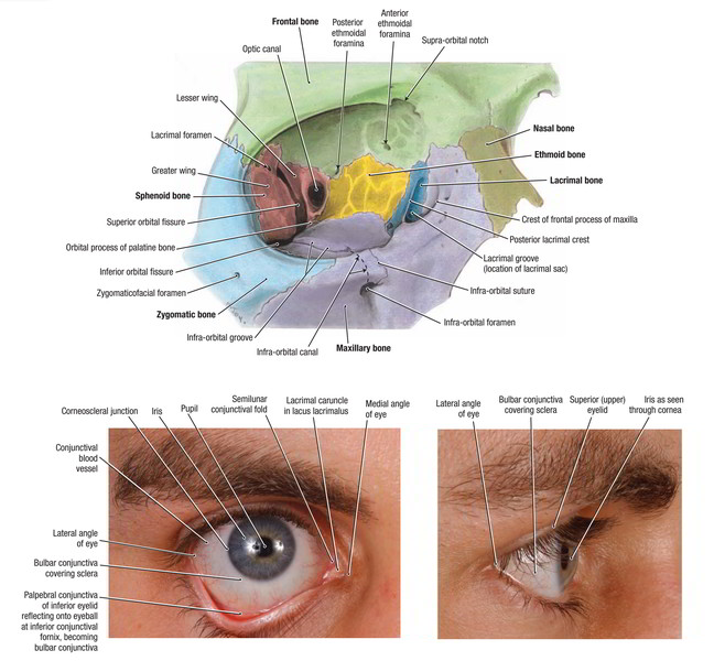
Palpebral Fissure
When the eye is open, the eyelids are divided by an elliptical fissure referred to as palpebral fissure.
Features Viewed Via Palpebral Fissure
- Lacus lacrimalis, a small triangular space in the medial part with reddish fleshy appearing elevation in its center referred to as lacrimal caruncle.
- Plica semilunaris, a small curved fold of conjunctiva immediately lateral to lacrimal caruncle. It represents nictitating membrane of lower creatures.
- Cornea and pupil, present in its centre. Notice that upper one seventh of the cornea is overlapped by the upper eyelid.
- Sclera of eye ball, viewed as white regions on each side of cornea.
Development
Above and below the cornea, the eyelids grow as folds of skin, which come together and stick along their borders during the third month of intrauterine life. The space between the folds and the cornea is the conjunctival sac.
When the eyelids separate, the palpebral fissure is established.
The orbital septum grows from mesodermal core in the skin folds. When the eyelids are closed, the orbital septum from orbital margin to the eyelids creates an entire diaphragm of the orbital cavity; the medial part of orbital septum enters behind the lacrimal sac and is connected to the lacrimal bone. This connection creates sharp posterior lacrimal crest.
Conjunctiva
The conjunctiva is a thin transparent mucus membrane, which lines the inner surfaces of eyelids (palpebral conjunctiva) and the front of sclera and cornea of eyeball (bulbar conjunctiva). The potential space between eyelids and eyeball when eyes are closed is named conjunctival sac. The lines of reflexion between palpebral and bulbar conjunctiva above and below create the superior and inferior fornices, respectively.
The palpebral conjunctiva is highly vascular and firmly adherent to the tarsal plates. On the flip side, bulbar conjunctiva is loose over the sclera but firmly adherent to the cornea creating its anterior epithelium (the corneal epithelium). The conjunctiva includes mucus-secreting goblet cells.
Conjunctival Fluid
The conjunctival sac is filled up with 3 pictures of fluid from inside outwards these are:
- Watery from lacrimal fluids.
- Mucus from conjunctiva.
- Fatty from tarsal glands.
The flashing movements of eyelids make these pictures moisten cornea and help drain the conjunctival fluid into nasal cavity.
Clinical Significance
Ptosis: It’s the drooping of the upper eyelid as a result of paralysis of levator palpebrae superioris following lesion of occulomotor nerve, which supplies this muscle.
Stye (hordeolum): It’s the suppurative inflammation of the Zeis gland In this state, the gland is swollen, difficult and debilitating. The pus points near the base of the cilia, therefore can be easily emptied by plucking the cilia.
Chalazion: It’s the inflammation of the tarsal (Meibomian gland). It causes localized swelling on the inner aspect of the eyelid.
Blepharitis: It’s the inflammation of the eyelids typically demanding eyelid margins.
Surgical operations on the lacrimal sac, consequently, are consistently anterior (outside) to the orbital cavity suitable because orbital septum enters behind the lacrimal sac to obtain connection on the posterior lacrimal crest.
The conholesation and thickening of orbital septum inside the growing eyelids creates the tarsal plates.
The inflammation of conjunctiva (conjunctivitis) because of infection or allergy is just one of the commonest diseases of the eye. Trachoma is a granular conjunctivitis caused by trachoma virus. It’s infectious and considered as the commonest cause of blindness in India.
It’s common for doctor to examine palpebral conjunctiva for anemia and bulbar conjunctiva for jaundice.
Nerve Supply
The conjunctiva is supplied by the following nerves:
- Palpebral conjunctiva of upper eyelid and ocular conjunctiva- by ophthalmic nerve.
- Palpebral conjunctiva of lower eyelid- by maxillary nerve.
Arterial Supply
The conjunctiva is supplied by palpebral and anterior ciliary arteries originated from ophthalmic artery.
Venous Drainage
The venous blood from palpebral conjunctiva is drained into facial vein while from ocular conjunctiva into ophthalmic veins.
Lymph Drainage
The lymph from conjunctiva is drained into preauricular lymph nodes.

 (57 votes, average: 4.67 out of 5)