The cancers originating in this region have predictable patterns of spread via the chains of lymph nodes in the neck, which help the surgeons to eliminate the desirable lymph nodes, the understanding of lymphatic drainage of the head and neck is vitally essential since. The lymph nodes and other lymphoid tissues in the head and neck are analyzed by doctors in day to day practice, which are frequently inflamed and generate swellings. Either (a) directly from the tissues or (b) indirectly after going through the outlying groups of lymph nodes, all the lymph from the region of head and neck drains into deep cervical lymph nodes.
On the right side drains into the right lym-phatic duct and on the left side into the thoracic duct the efferents from deep cervical nodes create the jugular trunk. Typically the right lymphatic duct and thoracic duct empty into the junction of the subclavian and internal jugular veins on their various sides.
Lymph Nodes
Out of absolute 800 lymph nodes within the body, about 300 lymph nodes are found in the region of the head and neck only. The lymph nodes in the region of the head and neck are generally classified into 2 groups: peripheral and concluding
Peripheral Lymph Nodes
Peripheral lymph nodes (also termed outlying lymph nodes). They can be generally seen in groups, which are ordered in outer and inner circles:
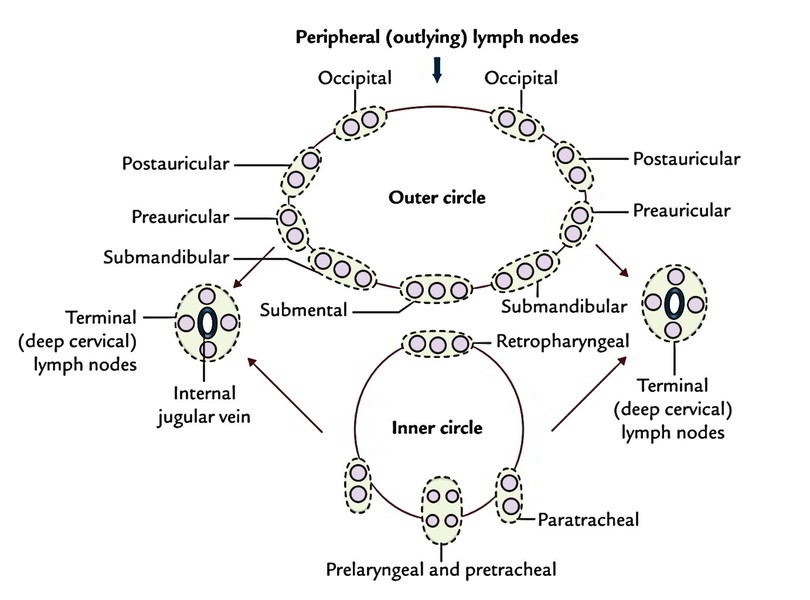
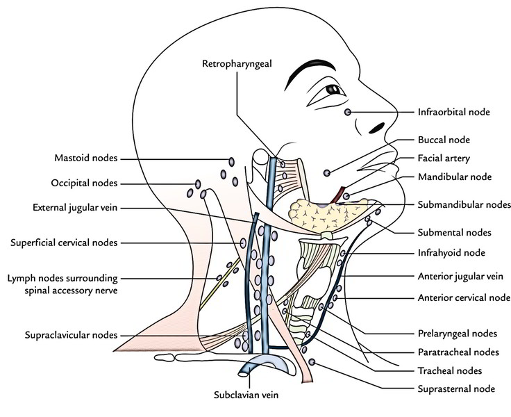
Outer circle: It’s created by lymph node groups, which create the pericervical or cervical collar atthe junction of the head and neck (craniocervical junction) and stretches from chin in front to the occiput behind. They contain submental, submandibular, superficial parotid (preauricular), mastoid (postauricular) and occipital nodes.
Outlying extensions of lymph node groups of pericervical collar:
Facial nodes: These are extensions of submandibular nodes and contain:
- a small buccal node being located on the lateral surface of the buccinator along the facial vein.
- a small mandibular node that’s often present where facial vessels cross the lower border of the mandible.
- a small infraorbital node being located just below the orbit.
Superficial cervical nodes: they’re situated superficial to sternomastoid (upper part) along the external jugular vein. All these really are the extensions of parotid nodes.
Anterior cervical nodes: they’re situated along the anterior jugular vein. 1 member of the group often is located in the suprasternal space (suprasternal node). They’re extensions of submental lymph nodes.
Inner circle: The inner circle is composed by subsequent lymph node groups, which is located deep to the investing layer of deep cervical fascia :
- Infrahyoid nodes: These be located in front of thyrohyoid membrane.
- Prelaryngeal nodes: These are situated in front of the conus elasticus or cricothyroid membrane.
- Pretracheal lymph nodes: These is located in front of trachea below the isthmus of thyroid gland
- Paratracheal nodes: These nodes flank the trachea and esophagus on each side along the recurrent laryngeal nerves.
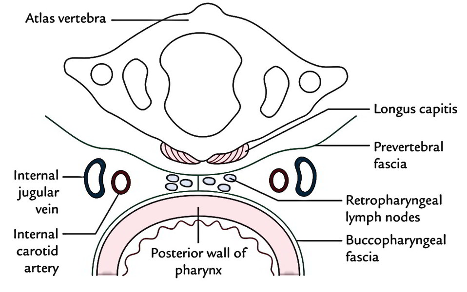
- Retropharyngeal lymph node: These be located posterior to pharynx and in front of prevertebral fascia in the retropharyngeal space .
Deep to inner circle, there’s a submucosal ring of aggregated masses of lymphoid tissue referred to as tonsils, which surround the commencement of air and food passages. These collectively represent the Waldeyer’s lymphatic ring . The lymph from lymphoid tissue of the ring.
Terminal Lymph Nodes
All these are deep cervical lymph nodes that be located along and around the internal jugular vein, some inside the carotid sheath and some on the surface of the sheath, under cover of sternocleidomastoid.
For the ease of description deep cervical lymph nodes are split into upper and lower groups (superior and inferior deep cervical nodes), though there’s no clear difference between them:
- Superior group of deep cervical lymph nodes: They are located above the omohyoid. 1 lymph node of the group is situated below the posterior belly of digastric between the angle of the mandible and anterior border of the sternocleidomastoid in the triangle created by posterior belly of digastric, facial vein and internal jugular vein. It’s referred to as jugulodigastric node.
It empties the lymph mainly from the palatine tonsil. Hence it’s also referred to as lymph node of the tonsil. When enlarged because of pathology in the palatine tonsil, it’s easily palpable behind and below the angle of the mandible.
- Lower group of deep cervical lymph nodes: 1 of thelymph nodes of the group is located above the intermediate tendon of omohyoid posterior to the internal jugular vein. It’s referred to as jugulo-omohyoid lymph node. Since this lymph node drains lymph mainly from the tongue, it’s called lymph node of the tongue. This node is located deep to sternocleidomastoid and for that reason, can be palpated only if enlarged significantly.
Some nodes of the group extend into the supraclavicular fossa and are related to brachial plexus and subclavian vessels. All these are called supraclavicular lymph nodes (Virchow’s lymph nodes). The left supraclavicular lymph nodes are medically essential since they’re common site of metastasis from malignant disease (cancer) of the stomach. The testicular and esophageal cancers can also metastasize in these nodes. The Virchow’s lymph nodes are frequently palpable in cancer stomach. 1 or 2 lymph nodes of the group is located in contact with accessory nerve at a higher level in the posterior triangle.
Clinical Significance
Surgical Neck Dissection
The cancers appearing in the head and neck region from structures like nasopharynx, paranasal air sinuses, oral cavity, oropharynx, larynx and thyroid gland have predictable patterns of spread via the chains of lymph nodes in the neck. When functioning to eliminate malignant lesion in this region, it’s vitally essential to understand these patterns of spread. The surgeons classify lymph nodes in neck into these levels:
Level I nodes are in the submental and submandibular triangles.
- Level II nodes are located around the upper portion of internal jugular vein and upper part of spinal accessory nerve. They go from the base of the skull to the bifurcation of the common carotid artery or the hyoid bone.
- Level III nodes are located around the middle third of the internal jugular vein and go from inferior border of level II to the intermediate tendon of omohyoid (cricoid cartilage).
- Level IV nodes are located around the lower third of the internal jugular vein and go from lower border of level III to the clavicle. It also contains supraclavicular lymph nodes.
- Level V nodes are in the posterior triangle of the neck, related to the spinal accessory nerve.
- Level VI nodes are nodes enclosing the midline visceral structures and contain the pretracheal and paratracheal nodes.
- Level VII nodes are in the superior mediastinum. Knowing which levels of nodes will probably participate in the metastatic spread of a specific cancer, a proper nodal clearance is undertaken.
The ancient radical neck dissection entails the removal of level I to V nodes and removal of the sternocleidomastoid muscle, internal jugular vein and spinal accessory nerve.
The modified radical neck dissection (also referred to as functional neck dissection) affects the removal of level I to V nodes but spares either or all of sternocleidomastoid muscle, internal jugular vein and spinal accessory nerve.
The selective neck dissection affects some but not level I to V nodes.

 (48 votes, average: 4.58 out of 5)