Muscles of The Gluteal Region is the area that covers region of the rear and the side, of the lateral half of the pelvis. It covers the iliac crest from above to the gluteal fold from below. It goes laterally up to a virtual line converging the anterior superior iliac spine to the anterior edge of the higher trochanter and medially goes upto mid-dorsal line and natal cleft. It is the most common site of administering intra-muscular injections.
The muscles of the gluteal region are split into 2 groups- major and minor.
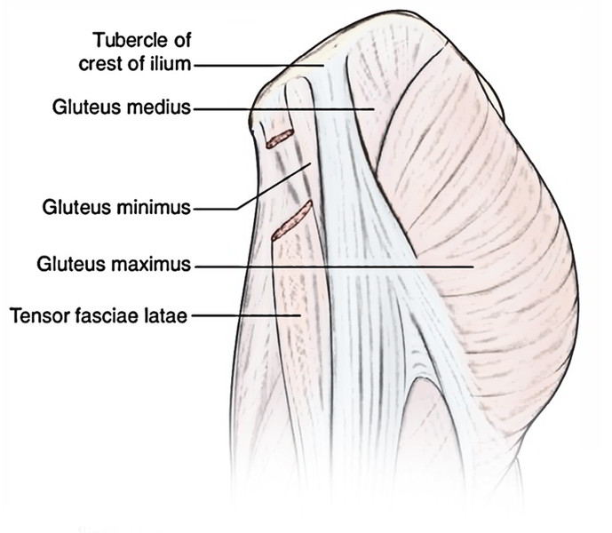
The major muscles of gluteal region are bigger in size and set superficially. They’re primarily extensor, abductors, and medial rotators of the thigh.
All these are 4 in number as given below:
- Gluteus maximus.
- Gluteus medius.
- Gluteus minimus.
- Tensor fasciae latae.
The minor muscles of gluteal region are smaller in size and set deeply under cover of the lower part of the gluteus maximus. They may be lateral rotators of the thigh and help stabilize the hip joint.

All these are 6 in number as follows:
- Piriformis.
- Superior and inferior gemelli.
- Obturator internus.
- Quadratus femoris.
- Obturator externus.
Attachments, Nerve Supply, and chief Actions of the Muscles of the gluteal region.
| Muscle | Origin | Insertion | Nerve supply | Actions |
|---|---|---|---|---|
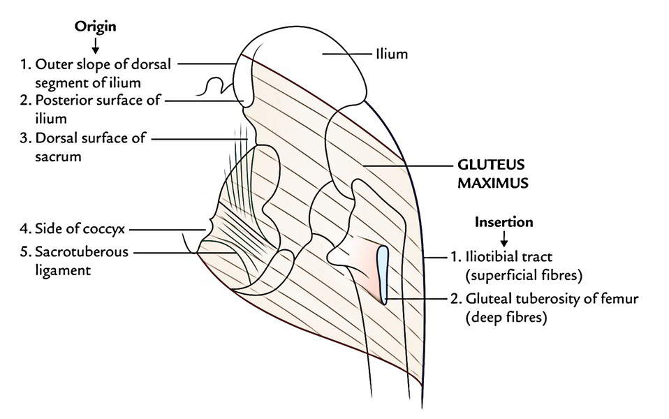
| Gluteus maximus (quadrilateral muscle) | 1. Gluteal surface of the ilium behind posterior gluteal line 2. Outer slope of the dorsal segment of ilium 3. Dorsal surfaces of the sacrum and ilium 4. Sacrotuberous ligament | 1. 3/4th of the muscle into the iliotibial tract 2. 1/4th of the muscle into the gluteal tuberosity | Inferior gluteal nerve (L5; S1, S2) | 1. Chief extensor of the hip joint 2. Assists in getting up from sitting position |
| Gluteus medius (fan-shaped muscle) | Gluteal surface of the ilium between anterior and posterior gluteal lines | Oblique ridge on the lateral surface of the greater trochanter | Superior gluteal nerve (L5; S1) | 1. Abductor of the hip joint 2. Prevents the sagging of pelvis on the unsupported side |
| Gluteus minimus (fan-shaped muscle) | Gluteal surfaces of the ilium between anterior and inferior gluteal lines | Ridge on the lateral part of the anterior and inferior gluteal lines trochanter | ||
| Tensor fasciae latae (fusiform muscle) | Outer lip of the anterior part of iliac crest (from ASIS to tubercle) | Iliotibial tract | Superior gluteal nerve | Supports the femur on tibia during standing position |
| Piriformis | Piriformis | Piriformis | Piriformis | Piriformis |
| (pear-shaped muscle, Latin pirum = pear) | Pelvic surface of the middle three pieces of sacrum by three digitations | Apex/tip of greater trochanter | Ventral rami of S1, S2 | Lateral rotator of the thigh at hip joint |
| Gemellus superior | Posterior surface of the ischial spine | Medial surface of greater trochanter along with tendon of obturator internus | Nerve to obturator internus (L5; S1, S2) | Lateral rotator of the thigh at hip joint |
| Gemellus inferior | 1. Upper part of the ischial tuberosity 2. Lower part of greater sciatic notch | Same as that of gemellus superior | Nerve to quadratus femoris (L4; L5, S1) | Lateral rotator of the thigh at hip joint |
| Obturator internus (fan-shaped muscle) | Pelvic surface of the obturator membrane and surrounding bones | Medial surface of greater trochanter of femur in front of trochanteric fossa | Nerve to obturator internus (L5; S1) | Lateral rotator of the thigh at hip joint |
| Quadratus femoris (quadrilateral muscle) | Lateral border of the ischial tuberosity | Quadrate tubercle on the intertrochanteric crest and area below it | Nerve to quadratus femoris (L5; S1) | Lateral rotator of the thigh at hip joint |
Major Muscles of Gluteal Region
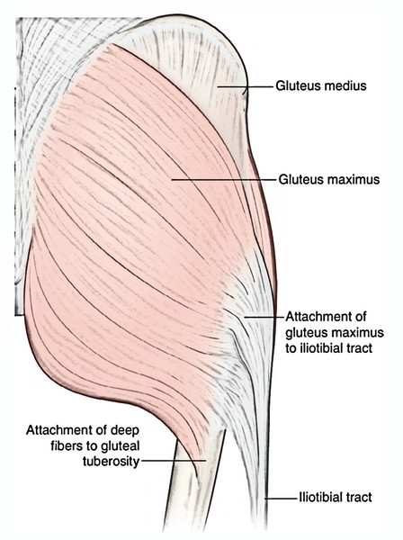
Gluteus Maximus
The gluteus maximus is the largest, most coarsely fibred, and most superficial gluteal muscle. It’s quadrilateral in shape and covers all of the other gluteal muscles with the exception of the anterosuperior part of the gluteus medius- the common site for intramuscular injections.
Origin
It originates from:
- Posterior part of the gluteal surface of ilium above and behind the posterior gluteal line.
- Outer sloping surface of the dorsal section of iliac crest.
- Aponeurosis of erector spinae muscle.
- Dorsal surfaces of the lower part of sacrum and adjoining part of the coccyx.
- Sacrotuberous ligament.
Insertion
The insertion takes place as follows:
- Quarter of the muscle (deep fibres of the lower part) is added into the gluteal tuberosity of femur.
- Three fourth of the muscle (superficial and deep fibres of the upper part) is added into the iliotibial tract, which in turn adds itself on the lateral condyle of the tibia.
Nerve Supply
The gluteus maximus is supplied by the inferior gluteal nerve (L5; S1, S2).
Actions
- The gluteus maximus is the main extensor of the hip joint during standing-up from sitting position and scaling upstairs.
- It plays an extremely significant part in rising from a sitting position and keeping the erect stance.
- Structures under cover of the gluteus maximus muscle.
| Muscles | Vessels | Nerves | Joints and ligaments | Bursae |
|---|---|---|---|---|
| All the muscles of the gluteal region except tensor fasciae latae | Superior and inferior gluteal vessels | Superior and inferior gluteal nerves | Hip joint | Trochanteric |
| bursa | bursa | bursa | bursa | bursa |
| Reflected head of the rectus femoris | Internal pudendal vessels | Sciatic nerve | Sacroiliac joint | Ischial bursa (occasional) |
| Origin of hamstrings | Trochanteric arterial anastomosis | Posterior cutaneous nerve of the thigh | Sacrotuberous | Origin of hamstrings |
| ligament | Gluteo-femoral | ligament | Gluteo-femoral | ligament |
| bursa | bursa | bursa | bursa | bursa |
| Insertion of the upper fibres of the adductor magnus | Cruciate arterial anastomosis | 1. Nerve to quadratus femoris 2. Pudendal nerve 3. Nerve to obturator internus 4. Perforating cutaneous nerve | 1. Sacrospinous ligament 2. Ischiofemoral ligament |
|
Gluteus Medius
The gluteus medius is a fan-shaped muscle. Its posterior one-third is deep and covered by the gluteus maximus while its anterior two-third is superficial and not covered by the gluteus maximus. Consequently, the intramuscular injection ought to be ideally supplied in this part.
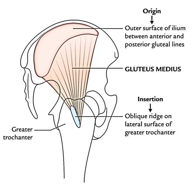
Origin
Gluteus medius originates from the gluteal surface of ilium between the anterior and posterior gluteal lines and gluteal aponeurosis.
Insertion
The muscle fibres converge downward, forward, and laterally to create a flat tendon that is added on to the oblique ridge on the lateral surface of the higher trochanter. The oblique ridge runs downward and forward from the tip of the higher trochanter.
- Between the tendon of gluteus medius and lateral surface of greater trochanter is located a bursa- the trochanteric bursa of gluteus medius.
Nerve Supply
The Nerve Supply is by the superior gluteal nerve.
Action
It’s the chief abductor of the hip joint.
Gluteus Minimus
The gluteus minimus is also a fan-shaped muscle and is located underneath the gluteus medius.
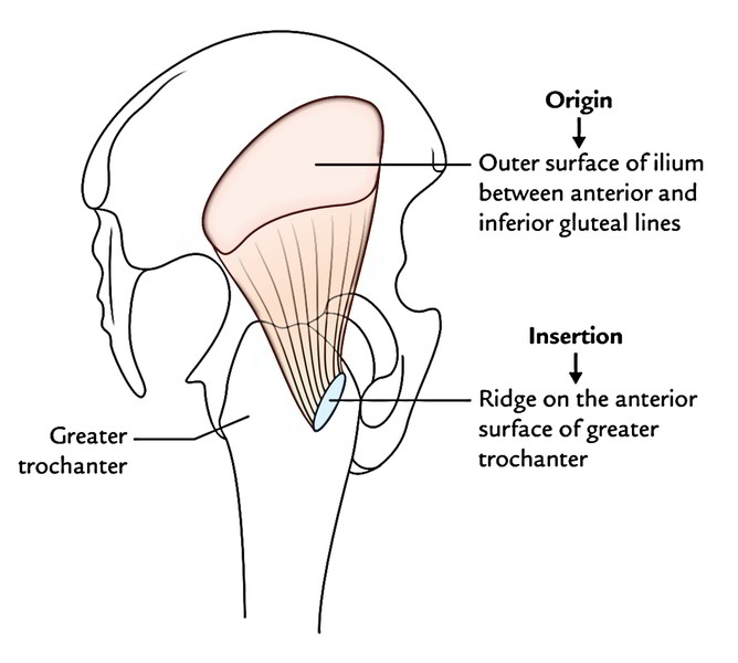
Origin
It appears from the gluteal surface of ilium between the anterior and inferior gluteal lines.
Insertion
The muscle fibres converge downward and somewhat laterally to create a tendon, that is added on to the ridge in the lateral part of the anterior surface of the higher trochanter.
- Between the tendon of gluteus minimus and the anterior surface of greater trochanter is located a bursa- the trochanteric bursa of gluteus minimus.
Nerve Supply
The gluteus minimus is supplied by the superior gluteal nerve.
Action
It’s the abductor of the hip joint.
Abductor mechanism of the hip joint
This mechanism includes 3 parts: (a) power (P) supplied by the gluteus medius and minimus, (b) fulcrum (F) supplied by the hip joint, and (c) weight (W) by the head and neck of thefemur. If any of these parts is deranged, the gluteal lax happens.
Clinical significance
Trendelenburg’s hint: Acting from below, both the gluteus medius and minimus keep the unsupported side of the pelvis from sagging during walking and so keep the horizontal level of the pelvis supplied the hip joint and neck-shaft angle of the femur are normal.
When the gluteus medius and minimus of 1 side are paralyzed as a result of injury of the superior gluteal nerve. The pelvis sags on the healthy side if that foot is off the earth. Consequently, the individual walks with a lurching gait. This is medically called Trendelenburg’s indication.
Tensor Fasciae Latae

The tensor fasciae latae muscle is the most anterior of the superfcial group of muscles in the gluteal region and overlies the gluteus minimus and the anterior part of the gluteus medius. The tensor fasciae latae originates fom the outer margin of the iliac crest from the anterior superior iliac spine to approximately the tuberculum of the iliac crest.
The muscle fibers descend to insert into the anterior aspect of the iliotibial tract of deep fascia, which runs down the lateral side of the thigh and attaches to the upper tibia. Like the gluteus maximus muscle, the tensor fasciae latae is enclosed within a compartment of the fascia lata.
The tensor fasciae latae stabilizes the knee in extension and, working with the gluteus maximus muscle on the iliotibial tract lateral to the greater trochanter, stabilizes the hip joint by holding the head of the femur in the acetabulum. It is innervated by the superior gluteal nerve
Minor Muscles of Gluteal Region
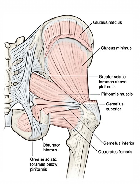
Piriformis
The piriformis muscle is the most superior of the deep group of muscles and is a muscle of the pelvic wall and of the gluteal region. It originates from between the anterior sacral foramina on the anterolateral surface of the sacrum and passes laterally and inferioriy through the greater sciatic foramen.
In the gluteal region, the piriformis passes posterior to the hip joint and attaches to a facet on the upper margin of the greater trochanter of the femur.
The piriformis externally rotates and abducts the femur at the hip joint and is innervated in the pelvic cavity by the nerve to the piriformis, which originates as branches from SI and S2 of the sacral plexus.
In addition to its action on the hip joint, the piriformis is an important landmark because it divides the greater sciatic foramen into two regions, one above and one below the piriformis. Vessels and nerves pass between the pelvis and gluteal region by passing through the greater sciatic foramen either above or below the piriformis.
Obturator internus
The obturator internus muscle, like the piriformis muscle, is a muscle of the pelvic wall and of the gluteal region. It is a flat fan-shaped muscle originating from the medial surface of the obturator membrane and adjacent bone of the obturator foramen. Because the pelvic floor attaches to a thickened band of fascia across the medial surface of the obturator internus. the obturator internus forms:
- the anterolateral wall of the pelvic cavity above the pelvic flow, and
- the lateral wall of the ischio-anal fossa in the perineum below the pelvic floor.
The muscle fibers of the obturator internus converge to form a tendon, which bends 90® around the ischium between the ischial spine and ischial tuberosity and passes through the lesser sciatic foramen to enter the gluteal region. The tendon then passes posteroinferiorly to the hip joint and attaches to the medial surface of the superior margin of the greater trochanter of the femur just inferior to the attachment of the piriformis muscle.
The obturator intern us laterally rotates and abducts the femur at the hip joint and is innervated by the nerve to the obturator internus.
Gemellus superior and inferior
The gemellus superior and inferior {gemdli is Latin for “twins”) are a pair of triangular muscles associated with the upper and lower margins of the obturator internus tendon:
- The base of the gemellus superior originates from the gluteal surface of the ischial spine.
- The base of the gemellus inferior originates from the upper gluteal and pelvic surfaces of the ischial tuberosity.
Fibers of the gemellus muscles attach along the length of the obturator internus tendon, and the apices of the two muscles insert with the tendon of the obturator internus on the greater trochanter of the femur.
The gemellus superior is innervated by the nerve to the obturator internus. and the gemellus inferior is innervated by the nerve to the quadratus femoris. The gemellus muscles act with the obturator internus muscle to laterally rotate and abduct the femur at the hip joint.
Quadratus femoris
The quadratus femoris muscle is the most inferior of the deep group of muscles in the gluteal region. It is a flat rectangular muscle below the obturator internus muscle and its associated gemellus muscles.
The quadratus femoris is attached at one end to a linear roughening on the lateral aspect of the ischium just anterior to the ischial tuberosity and at the other end to the quadrate tubercle on the intertrochanteric crest of the proximal femur.
The quadratus femoris laterally rotates the femur at the hip joint and is innervated by the nerve to the quadratus femoris.

 (48 votes, average: 4.63 out of 5)