The plantaris muscle is missing in 7-10% of the human population. The muscle belly on its own is short, while the tendon {is one of|is among} the {largest|biggest} in the body. Operating in combination with the soleus and gastrocnemius muscles to give a very little quantity of plantar flex ion of the calf, the plantaris is an accessory muscle.
Interactive Anatomical Interface
[AnteriorLegMuscles]
[PosteriorLegMuscles]
Relationship to Other Structures
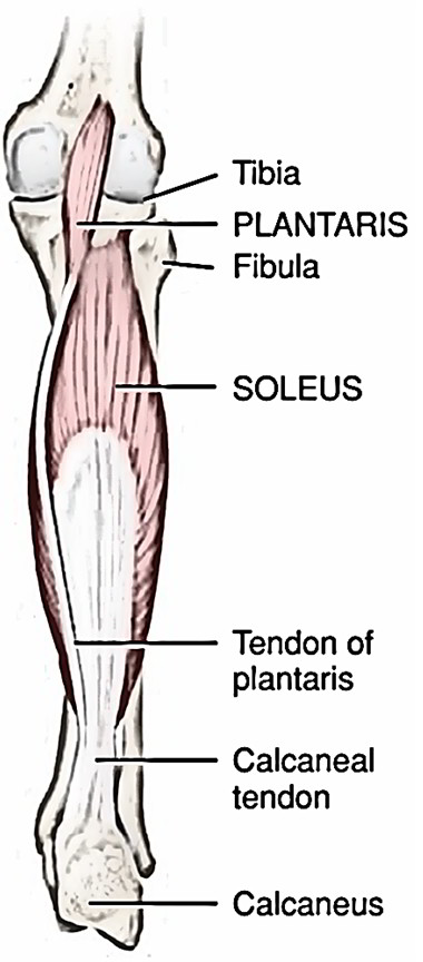
- Merely medial to the lateral head of the gastrocnemius and the distal tendon of the biceps femoris, the plantaris connects proximally.
- The plantaris is superficial to the popliteus here. Much of the belly of the plantaris is superficial in the posterior knee simply medial to the lateral head of the gastrocnemius.
- The remainder of the belly and the proximal V2 of the long distal tendon of the plantaris are sandwiched in between the gastrocnemius and the soleus. (The tendon is deep to the gastrocnemius and superficial to the soleus.).
- Precisely medial to the medial margin of the distal tendon of the gastrocnemius, the distal V2 of the long distal tendon is superficial.
- The plantaris is situated inside the superficial back line myofascial meridian.
Origin
The plantaris muscle has its musculotendinous junction simply distal to the level of the knee joint, and stems via the lateral femoral condyle, routes simultaneously the lower leg’s superficial posterior compartment.
Attachments
- It is placed into the calcaneus via the tendocalcaneus.
- The plantaris emerges from the lower-third of lateral supracondylar ridge of femur.
Insertion
- Its tendons are placed into the middle one-third of the posterior surface of the calcaneum medial to the attachment of tendocalcaneus and mixes with the medial margin of tendocalcaneus.
- The small spindle-shaped muscle body of the plantaris forms a thin tendon, and while comes down medially, deep to the lateral head of the gastrocnemius, which passes in between the gastrocnemius and soleus muscles and ultimately merges with the medial side of the calcaneal tendon near its connection to the calcaneus.
- It is usually called freshman’s nerve. The long slim tendon of plantaris is quickly misinterpreted by the first year medical trainees for a nerve.
Nerve Supply
Plantaris muscle is supplied by Tibial Nerve, S1, S2.
Arterial Supply
Sural branches of the Popliteal Artery (the extension of the Femoral Artery).
Actions
- It plantarflexes the foot at the ankle joint. Plantarflexion of the foot at the ankle joint is a crucial action throughout the gait cycle when pushing off the ground (toe-off stage) The plantaris crosses the ankle joint posteriorly (with its fibers running vertically in the sagittal plane).
- It bends the leg at the knee joint the plantaris crosses the knee joint posteriorly (with its fibers running vertically in the sagittal plane).
- The plantaris inverts the foot (more particularly, it inverts/supinates the calcaneus relative to the talus) at the subtalar joint. The plantaris crosses into the foot to connect onto the calcaneus medial to the axis of the subtalar joint.
- Subtalar supination likewise consists of medial rotation and plantarflexion of the foot, inversion is among the significant part of subtalar supination.
Clinical Examination
- With the patient supine with the leg extended at the knee joint, put palpating fingers on the proximal posterior leg, at the lateral border of the popliteal fossa (simply medial to the proximal connection of the lateral head of the gastrocnemius).
- Plantarflex the foot at the ankle joint and understanding of the constriction of the belly of the plantaris and ask the customer to actively bend the leg at the knee joint. (Distinguishing the plantaris via the lateral head of the gastrocnemius will be hard considered that the gastrocnemius will likewise contract with these two actions.).
Rate this Article: 
 (46 votes, average: 4.87 out of 5)
(46 votes, average: 4.87 out of 5)