Muscles of the posterior (flexor) compartment of the leg are divided into two groups, superficial and deep, separated by a layer of deep fascia. Generally, the muscles mainly plantarflex and invert the foot and flex the toes. The tibial nerve gives supply to all of them.
Superficial and deep muscles of the posterior compartment of the leg.
| Superficial muscles | Deep muscles |
|---|---|
| Gastrocnemius | Popliteus |
| Soleus | Flexor digitorum longus |
| Plantaris | Flexor hallucis longus |
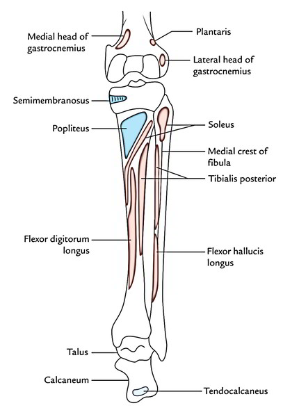
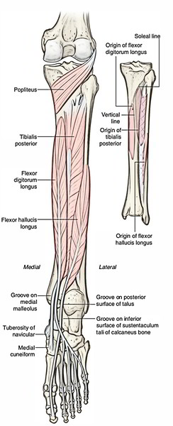
The bony attachments of the muscles of the back of the legare revealed in Figure below.
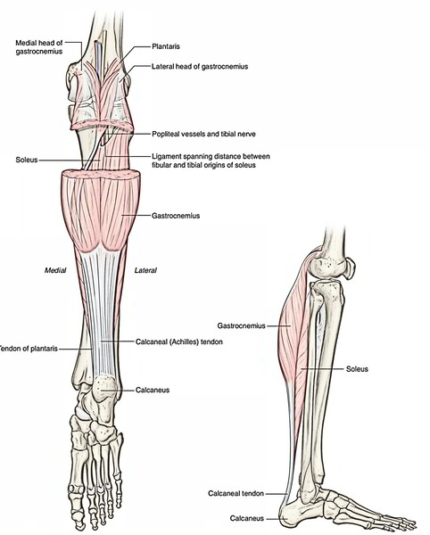
Superficial Muscles
The superfcial group of muscles in the posterior compartment of the leg comprises three muscles-the gastrocnemius, plantaris, and soleus, all of which insert onto the heel (calcaneus) of the foot and plantarflex the foot at the ankle joint. As a unit, these muscles are large and powerful because they propel the body forward off the planted foot during walking and can elevate the body upward onto the toes when standing. Two of the muscles (gastrocnemius and plantaris) originate on the distal end of the femur and can also flex the knee.
Origin, Insertion, Nerve Supply, and Actions of The Superficial Muscles of The Rear of The Leg
| Muscle | Origin | Insertion | Nerve supply | Actions |
|---|---|---|---|---|
| Gastrocnemius | 1. Lateral head from the lateral surface of the lateral condyle of the femur 2. Medial head from the popliteal surface of the femur and adjoining posterosuperior aspect of the medial condyle of the femur | Middle one-third of the posterior surface of the calcaneus via tendocalcaneus | Tibial nerve (S1, S2) | 1. Plantar flexion of the ankle when the knee is extended 2. Flexes the leg at the knee joint |
| Soleus | 1. Posterior aspect of the head and upper one-fourth of the posterior surface of the shaft of fibula 2. Soleal line and middle one- third of the medial border of the tibia 3. Tendinous soleal arch between the fibula and tibia | Middle one-third of the posterior surface of the calcaneus via tendocalcaneus | Tibial nerve via two branches (S1, S2) | 1. Plantar flexion of the ankle independent of position of the knee 2. Steadies the leg on the foot during standing |
Gastrocnemius
It’s the largest and most superficial muscle of the posterior compartment. It includes 2 heads- medial and lateral.
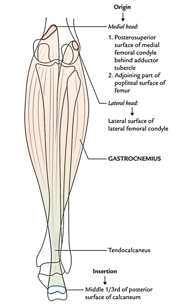
Origin
- Large medial head originates by a broad flat tendon from the posterosuperior aspect of the medial condyle of the femur supporting the adductor tubercle and adjoining part of the posterior (popliteal) surface of the shaft of the femur.
- Small lateral head originates by a broad flat tendon from the lateral surface of the lateral condyle of the femur above the lateral epicondyle and adjoining part of the lateral supracondylar line.
Insertion
- The fleshy midriffs of both heads descend and link in the middle of the leg to create broad thin aponeurotic tendon, which connects with the tendon of soleus, a short distance below the middle of the leg to create a long thick tendon- the tendocalcaneus (tendoachillis), that is added into the middle of the posterior surface of calcaneum.
Nerve Supply
Both heads are supplied by the tibial nerve in the popliteal fossa.
Actions
- It’s the main plantar flexor of the foot in the ankle when the knee is extended.
- It’s also a flexor of the knee.
- It gives fast movements of the foot during running and leaping.
Clinical significance
Tennis leg: It’s distressing calf injury where there’s rip or strain of the medial head of gastrocneminus at its musculotendinous junction because of overstretching. It typically appears in a tennis player who extends their gastrocnemius too much for a hard function.
Soleus
It’s a multipennate muscle being located deep to the gastrocnemius. It’s so termed due to its likeness to a sole/flat fish.

Origin
It’s a horseshoe-shaped origin from:
- Back of the head and posterior surface of the upper 1 fourth of the shaft of the fibula.
- Soleal line and middle 1/3rd of the medial border of the tibia.
- Tendinous arch between the tibia and fibula.
Insertion
The muscle fibres slope downward to create a gigantic abdomen, which provides rise to a tendon. The tendon of soleus fuses with that of gastrocnemius to create tendocalcaneus, that is added into the middle 1/3rd of the posterior surface of calcaneum.
Nerve Supply
It’s supplied by 2 branches of the tibial nerve:
- The first branch supplies the muscle in the popliteal fossa from its superficial surface.
- The 2nd branch supplies the muscle in the posterior compartment of the leg from its deep surface.
Actions
- It’s a plantar flexor of the foot in the ankle and steadies the leg on the foot during standing. It’s regarded as the “workhorse” of the plantar flexion.
- It’s slow acting but more strong than gastrocnemius. It functions as a bottom gear during wandering. (Note: Gastrocnemius functions as a top gear during running and leaping.).
Key Points
- The 2 heads of gastrocnemius and soleus together form the triceps surae muscle.
- A small sesamoid bone is seen in the tendon of origin of lateral head of gastrocnemius referred to as fabella.
- A small synovial bursa (Brodie’s bursa) is located deep to medial head to gastrocnemius.
- Tendocalcaneus (tendoachillis) is a conjoint tendon of insertion of gastrocnemius and soleus (triceps surae). It’s the thickest and strongest tendon in the entire body and is about 15 cm long. It functions as a prime mover of plantar flexion of the foot in the ankle joint.
Clinical Testing
To examine gastrocnemius and soleus together (triceps surae), request the patient to stand on the toes or plantar flex the foot. If normal the tendocalcaneus can be viewed and palpated above the heel.
Clinical significance
Calf muscle pump and peripheral heart: The gastrocnemius and soleus jointly make a ‘calf muscle pump which eases the venous return from the lower limb.
The soleus muscle is regarded as the peripheral heart as it houses large venous sinuses referred to as soleal sinuses which convey with all the superficial veins by perforating veins and with deep veins directly.
Consequently, contraction of soleus helps in sucking the blood from superficial veins and propelling it into deep veins.
Plantaris
It’s a small muscle with a short gut and long slim tendon. It is located between the gastrocnemius and soleus.
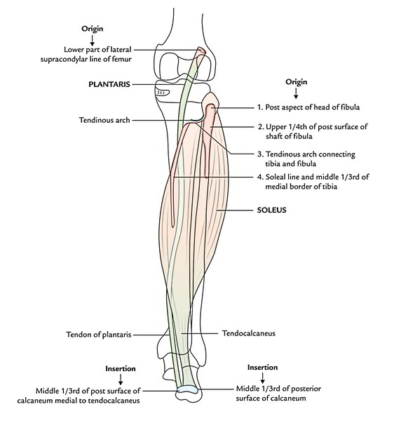
Origin.
It appears from the lower 1/3rd of the lateral supracondylar line and the adjoining part of the oblique popliteal ligament.
Insertion.
Its tendon combines with all the medial margin of tendo calcaneus and fit into the middle 1/3rd of the posterior surface of the calcaneum medial to the connection of tendo calcaneus.
The long slim tendon of plantaris is easily mistaken by the very first year medical students for a nerve; thus, it’s frequently called freshman’s nerve.
Clinical significance
Tendon grafting: The plantaris is a vestigial muscle in human beings and is absent in 5-10% of individuals. It’s trivial job either as flexor of the knee or as plantar flexor of the ankle; thus, the tendon of plantaris is utilised for grafting (example, reconstructive surgery of the tendons of the hand).
Deep Muscles
There are four muscles in the deep posterior compartment of the leg – the popliteus, flexor hallucis longus, flexor digitorum longus, and tibialis posterior. The popliteus muscle acts on the knee, whereas the other three muscles act mainly on the foot.
Origin, insertion, Nerve Supply, and Actions of the deep muscles of the back of the leg.
| Muscle | Origin | Tendon | Insertion | Nerve supply | Actions |
|---|---|---|---|---|---|
| Popliteus | Anterior part of the popliteal groove on the lateral condyle of the femur | Its tendon of origin is 1 inch long, and intracapsular but extrasynovial | Medial two-third of the triangular area above the soleal line, on the posterior surface of the tibia | Tibial nerve (L4, L5; S1) | Unlocks the locked knee by rotating the femur laterally during initial stages of flexion of the knee |
| Flexor digitorum longus (FDL) | 1. Upper two-third of the medial part of the posterior surface of the tibia below the soleal line. 2. Fascia covering the tibialis posterior. | Its tendon: 1. Crosses superficially to the tendon of tibialis posterior in the lower part of the leg and tendon of FHL in the sole. 2. Subdivides into four tendons for lateral four toes, accessorius | Plantar surface of the base of distal phalanges of the lateral four toes. | Tibial nerve (S2, S3) | Its tendon: 1. Plantar flexes lateral four toes. 2. Plantar flexes ankle. 3. Maintains the longitudinal arches of the foot. |
| Flexor hallucis longus (FHL) | 1. Lower three-fourth of the posterior surface of the shaft of the fibula, behind the medial crest.2. Adjoining part of the posterior interosseous membrane | Its tendon: 1. Grooves two bones, talus between its medial and posterior tubercles and the calcaneum underneath sustentaculum tali. 2. In the sole it is crossed superficially by the tendon of FDL. | Plantar surface of the base of distal phalanx of great | Tibial nerve (S2, S3) | 1. Plantar flexes great toe. 2. Plantar flexes the ankle weakly. 3. Supports the medial longitudinal arch of the foot |
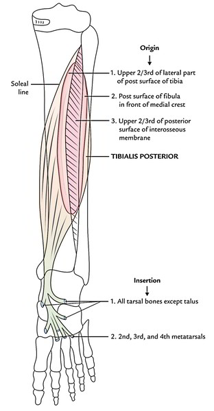
| Tibialis posterior | 1. Upper 2/3rd of the lateral part of the posterior surface of tibia below the soleal line. 2. Posterior surface of the fibula in front of the medial crest. 3.Upper two-third of the posterior surface of the interosseous membrane | Its tendon: 1. Crosses superficially by the tendon of FDL in the lower part of the leg. 2. Grooves back of the medial malleolus. 3. Passes deep to flexor retinaculum and superficially to the deltoid ligament | 1. Chiefly on tuberosity of the navicular bone. 2. Slips pass to all tarsals, except talus, and bases 2nd to 4th metatarsals | Tibial nerve (L4, L5) | 1.Invertor of the foot. 2.Maintains the medial and longitudinal arches of the foot |
Popliteus
It’s a thin, flat, triangular muscle, which forms the inferior part of the floor of the popliteal fossa.
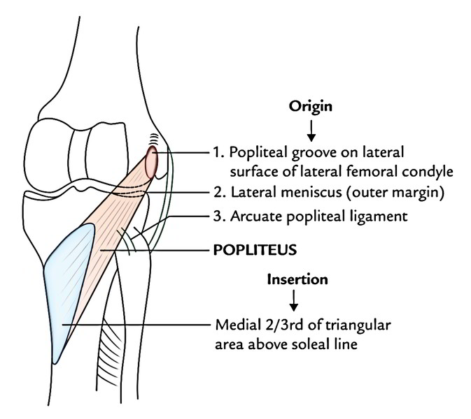
Origin
Its origin is intracapsular but extra synovial. It originates by a tendon from:
- The deep anterior part of the groove (popliteal groove) on the lateral surface of the lateral condyle of the femur. (Notice: The posterior part of the groove is inhabited by the tendon of popliteus in total flexion.).
- Arcuate popliteal ligament.
- Outer margin of the lateral meniscus.
Insertion
The tendon enters downward and medially and flares out to create the fleshy part, that is added into:
- The medial 2-3rd of the triangular area above the soleal line on the posterior outermost layer of the tibia.
- The popliteal fascia.
Nerve Supply
The popliteus is provided by a branch of tibial nerve. It originates in the popliteal fossa, crosses across the superficial aspect of the muscle, winds round its lower border, and provides the muscle from its deeper aspect.
Actions
- It unlocks the locked knee by rotating the femur laterally during first phases of flexion of the knee.
- It pulls the lateral meniscus backwards and keeps it from being trapped and crushed between the condyles of the femur and tibia.
- It bends the knee during couching.
Flexor hallucis longus
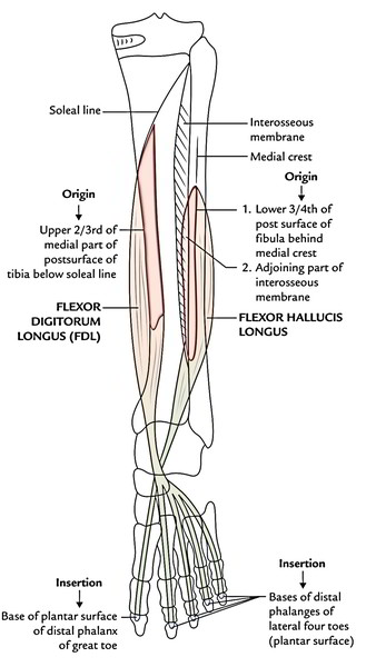
The flexor hallucis longus muscle originates on the lateral side of the posterior compartment of the leg and inserts into the plantar surface of the great toe on the medial side of the foot. It arises mainly from the lower 2/3rd of the posterior surface of the fibula and adjacent interosseous membrane.
The muscle fibers of the flexor hallucis longus converge inferiorly to form a large cord-like tendon, which passes behind the distal head of the tibia and then slips into a distinct groove on the posterior surface of the adjacent tarsal bone (talus) of the foot. The tendon curves anteriorly first under the talus and then under a shelf of bone (the sustentaculum tali), which projects medially from the calcaneus, and then continues anteriorly through the sole of the foot to insert on the inferior surface of the base of the distal phalanx of the great toe.
The flexor hallucis longus flexes the great toe. It is particularly active during the toe-off phase of walking when the body is propelled forward off the stance leg and the great toe is the last part of the foot to leave the ground. It can also contribute to plantarflexion of the foot at the ankle joint and is innervated by the tibial nerve.
Flexor digitorum longus
The flexor digitorum longus muscle originates on the medial side of the posterior compartment of the leg and inserts into the lateral four digits of the foot. It arises mainly from the medial side of the posterior surface of the tibia inferior to the soleal line.
The flexor digitorum longus descends in the leg and forms a tendon, which crosses posterior to the tendon of the tibialis posterior muscle near the ankle joint. The tendon continues inferiorly in a shallow groove behind the medial malleolus and then swings forward to enter the sole of the foot. It crosses inferior to the tendon of the flexor hallucis longus muscle to reach the medial side of the foot and then divides into four tendons, which insert on the plantar surfaces of the bases of the distal phalanges of digits II to V.
The flexor digitorum longus flexes the lateral four toes. It is involved with gripping the ground during walking and propelling the body forward off the toes at the end of the stance phase of gait. It is innervated by the tibial nerve.
Tibialis posterior
The tibialis posterior muscle originates from the interosseous membrane and the adjacent posterior surfaces of the tibia and fibula. It lies between and is overlapped by the flexor digitorum longus and the flexor halluces longus muscles.

 (49 votes, average: 4.56 out of 5)