Sole of the foot is the bottommost region of foot where human foot comes in contact with the earth. in various ways the structure of foot and palm is comparable and similar to the palm of human hand. The major difference between foot and palm is functional differences that they have. The hand is an organ capable of clasping while the foot is meant for transfer of body weight and movement of body. There are different parts of foot that are in contact with the earth at different type of activities like running, walking and standing.
Skin
The skin of the sole presents the following features:
- It’s thick and hairless.
- It’s securely bound to the underlying deep fascia (plantar aponeurosis) by the many fibrous bands.
- It’s creased at the ssites of skin movement.
- It includes large number of sweat glands.
The above features boost the efficiency of grip of the sole on the earth.
- The skin over the major weight bearing regions of the sole- the heel, lateral margin, and ball of the foot- is quite thick.
Superficial Fascia
The superficial fascia is mainly composed of subcutaneous fat in the meshwork of fibrous septa, which anchor the skin together with the underlying deep fascia. The superficial fascia is thick and dense over the weight bearing points to supply fibrofatty pillows at these ssites (example, posterior tubercles of the calcaneum, metatarsal heads, and pulps of the digits).
Cutaneous Nerves
The skin of the sole of the foot is supplied by 3 cutaneous nerves, which originate directly or indirectly from the tibial nerve.
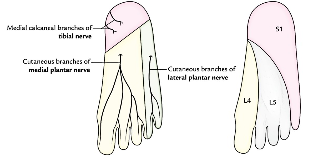
The regions supplied by the 3 nerves nearly correspond to the 3 dermatomes of the sole:
- Medial calcaneal branches: They originate directly from the tibial nerve and supply skin over the posterior and medial portions of the sole- the weight bearing portion of the heel. The corresponding dermatome is S1.
- Cutaneous branches of the medial plantar nerve: They supply the skin over the bigger anteromedial portion of the sole and medial 3 digits. The corresponding dermatome is L4.
Cutaneous branches of the lateral plantar nerve: They supply the skin over the smaller anterolateral portion of the sole and lateral 1 digits. The corresponding dermatome is L5.
Deep Fascia
The deep fascia in the region of the sole is composed of 3 parts: central, medial, and lateral. The central part of the deep fascia is quite thick and named plantar aponeurosis. The medial and lateral parts are thin and referred to as medial and lateral plantar fasciae, respectively.
The deep fascia of the sole consists of dense bundles of collagen fibres that are arranged longitudinally in the plantar aponeurosis and transversely in the medial and lateral plantar fasciae.
The thick central part covers the flexor digitorum brevis. The thin medial and lateral parts cover the abductor hallucis and abductor digiti minimi, respectively.
In the region of toes, the deep fascia forms the deep transverse metatarsal ligaments and fibrous flexor sheaths.
Key Points
- The deep fascia in the sole is specialized to create 3 things: (a) plantar aponeurosis, (b) deep transverse metatarsal ligaments, and (c) fibrous flexor sheaths.
- According to some authorities, entire of the deep fascia of the sole is named plantar aponeurosis.
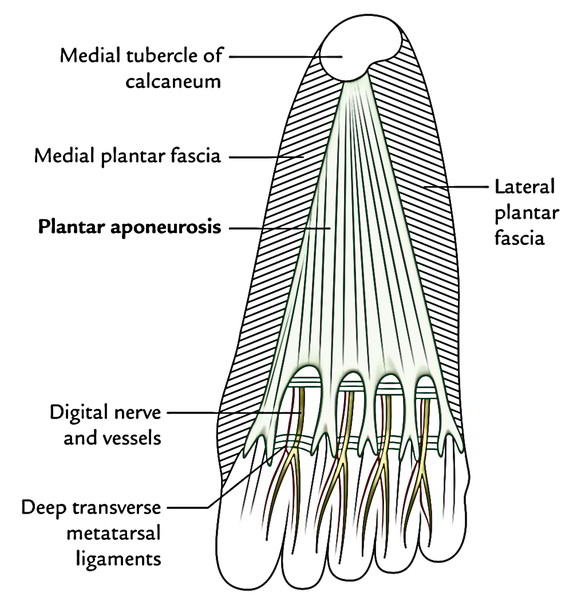
Plantar Aponeurosis
The plantar aponeurosis is the thickened central part of the deep fascia of the sole.
Features
- The plantar aponeurosis is triangular in shape and takes up the central area of the sole. The apex of the plantar aponeurosis is connected to the medial tubercle of calcaneum, proximal to the connection of the flexor digitorum brevis. The base of the plantar aponeurosis near the heads of the metatarsals breaks up into 5 groups, 1 for every toe. At the stage of division, the 5 processes are bound by the transverse fascial fibres. The digital nerves and vessels go through the separations between the processes.
- Every band divides opposite the metatarso-phalangeal joints into a superficial and a deep chemise. The superficial faux pas is connected to the dermis of the skin and fusion with the superficial transverse metatarsal ligaments. The deep chemise breaks up into 2 parts, which cover the flexor tendons, and mix together with the fibrous flexor sheaths and deep transverse metatarsal ligaments.
- From the medial and lateral margins of the aponeurosis, the medial and lateral vertical intermuscular septa pass deeply, and split the plantar muscles into 3 groups-medial, medium, and lateral. The narrower transverse septa originate from the vertical septa and break up the muscles of the sole into 4 layers.
- Morphologically the plantar aponeurosis represents the degenerated tendon of plantaris muscle, which has been divided by the enlarging heel during development.
Functions
The functions of the plantar aponeurosis are:
- It securely fixes the skin of the sole.
- It gives origin to the muscles of first layer of the sole.
- It shields the plantar nerve and vessels from compression.
- It helps to preserve the longitudinal arches of the foot by acting as tie beam.
Clinical Significance
Plantar fasciitis and calcaneal spur: The plantar aponeurosis is stretched during standing position. Consequently, splitting or inflammation (plantar fasciitis) frequently takes place in people who do a lot of standing or walking, viz. traffic police staff. It causes pain and tenderness in the sole of the foot particularly underneath the heel during standing. Continued episode of the plantar fasciitis results in calcification in the posterior connection of the plantar aponeurosis creating a calcaneal spur.
Deep Transverse Metatarsal Ligaments
All these are 4 short, flat bands of fibrous tissue, which attach the plantar ligaments of the adjoining metatarso-phalangeal joints. They’re linked dorsally to interossei, and ventrally to lumbricals and digital nerves and vessels.
Fibrous Flexor Sheaths
The inferior surface of every toe from the head of metatarsal to the base of distal phalanx is provided with a solid fibrous sheath originated from the deep fascia of the toes. It’s connected to the sides of the phalanges.
The proximal end of every sheath gets the deeper part of the slickness of plantar aponeurosis. The distal end of the sheath is closed and is connected to the base of the distal phalanx.
The sheath together with the inferior surfaces of the phalanges and interphalangeal joints creates a blind tunnel via which pass long flexor tendon/tendons of the toes.
Their structure and arrangement is quite similar to that of fibrous flexor sheaths of the fingers. They keep flexor tendons in position during flexion of the toes.
Muscles of The Sole of The Foot
There are 18 intrinsic muscles and 4 extrinsic tendons in the sole of the foot. The muscles of the sole are described in 4 layers from superficial to deep.
The muscles of the sole are primarily concerned with supporting the arches of the foot. The short and long muscles of the foot serve as synergists.
Neurovascular planes of the sole: There are 2 neurovascular planes between the muscle layers of the sole:
- Superficial neurovascular plane between the first and 2nd layers.
- Deep neurovascular plane between the 3rd and fourth layers.
In the superficial neurovascular plane is located the trunks of medial and lateral plantar nerves, and the arteries.
In the deep neurovascular plane is located the deep branches of the lateral plantar nerve and artery.
Muscle Layers of The Sole of The Foot
| Layer | Muscles | Features |
|---|---|---|
| First layer | 1. Flexor digitorum brevis. 2. Abductor hallucis. 3. Abductor digiti minimi | They cover whole of the sole |
| Second layer | 1. Flexor digitorum accessories Four lumbricals. 2. Two tendons (tendon of flexor digitorum longus and tendon of flexor hallucis longus) | Flexor digitorum accessorius and lumbricals are attached to the tendon of flexor digitorum longus |
| Third layer | 1. Flexor hallucis brevis. 2. Flexor digiti minimi brevis. 3. Adductor hallucis | 1. They are confined to the metatarsal region of the sole. 2. Two of these muscles act on the big toe and one on the little toe |
| Fourth layer | 1. Interossei (3 plantar interossei and 4 dorsal interossei). 2. Tendon of tibialis posterior. 3. Tendon of peroneus longus | They fill up the intermetatarsal spaces |
Origin, Insertion, Nerve Supply, and Actions
Mnemonic: Plantar interossei ADduct (PAD) the toes and originate from single metatarsal as unipennate muscles; on the other hand Dorsal interossei ABduct (DAB) the toes and originate from 2 metatarsals as bipennate muscles.
| Muscles | Origin | Belly/Tendon | Insertion | Nerve supply | Actions |
|---|---|---|---|---|---|
| • Flexor digitorum brevis (resembles flexor digitorum superficialis of the hand) | Medial tubercle of the calcaneum | 1.It forms 4 tendons for the lateral 4 toes. 2.Each tendon splits into two slips opposite the bases of proximal phalanges to allow passage of the long flexor tendon | Margins of the middle phalanges of lateral 4 toes | Medial plantar nerve (S2, S3) | Flexor of the lateral toes |
| • Abductor hallucis (lies along the medial border of the foot) | 1. Medial tubercle of the calcaneum. 2. Flexor retinaculum. | Its tendon fuses with medial portion of the tendon of flexor hallucis brevis for a common insertion | Medial side of the base of proximal phalanx of the big toe | Medial plantar nerve (S2, S3) | Flexion and abduction of the big toe |
| • Abductor digiti minimi (lies along the lateral border of the foot) | Medial and lateral tubercles of the calcaneum in a continuous line | Its tendon fuses with the tendon of flexor digiti minimi brevis for a common insertion | Lateral side of the base of proximal phalanx of the little toe | Lateral plantar nerve (S2, S3) | Flexion and abduction of the little toe |
| • Flexor digitorum accessorius | By two heads: 1. Medial head from the medial concave surface of the calcaneum and adjoining part of the medial tubercle. 2. Lateral head from the calcaneum in front of the lateral tubercle | The two heads unite at an acute angle | Tendon of the flexor digitorum longus (FDL) | Lateral plantar nerve (S2, S3) | 1. Straightens the pull of the long flexor tendons. 2. Assists the flexor digitorum longus in flexing the lateral 4 toes |
| • Lumbricals (4 in number and numbered from medial to lateral side) Tendon of flexor digitorum longus Tendon of flexor hallucis longus | From the tendons of the flexor digitorum longus | Tendons pass forwards on the medial sides of the metatarso-phalangeal joints of the lateral four toes | Into the extensor expansions and bases of proximal phalanges of lateral 4 toes | 1st by the medial plantar nerve, and 2nd, 3rd, and 4th by the lateral plantar nerve (S2, S3) | Extension of toes at the interphalangeal joints |
| Third Layer | |||||
| • Flexor hallucis brevis | It arises by a Y-shaped tendon: 1.Lateral limb from the medial part of the plantar surface of the cuboid bone. 2.Medial limb from the lateral cuneiform | Muscle belly splits in two parts. Each part gives rise to a tendon which contains a sesamoid bone near its insertion. The medial tendon blends with the abductor hallucis, and the lateral tendon blends with the adductor hallucis | Each side of the base of proximal phalanx of the big toe (1st digit) | Medial plantar nerve (S2, S3) | Flexion of the proximal phalanx of big toe |
| • Flexor digiti minimi brevis | Base of the 5th metatarsal bone | Forms narrow tendon which blends with the abductor digiti minimi | By a narrow tendon into the lateral side of the base of proximal phalanx of the little toe | Superficial branch of the lateral plantar nerve (S2, S3) | Flexes the proximal phalanx of the little toe |
| • Adductor hallucis | It arises by two heads: 1.Large oblique head from bases of the 2nd, 3rd, and 4th metatarsals. 2.Small transverse head from the plantar ligaments of the metatarso-phalangeal joints of 3rd, 4th, and 5th toes | The common tendon of both heads fuses with the lateral tendon of the flexor hallucis brevis | By the composite tendon on the lateral side of the base of proximal phalanx of the big toe | Deep branch of the lateral plantar nerve (S2, S3) | 1.Adduction of the big toe .2Maintains transverse arches of the foot. |
| • Dorsal interossei * (4 in number, and lies between the metatarsal bones) | They are bipennate and arises from the adjacent sides of the metatarsal bones (1-5) | Tendons are arranged on the abductor sides of the toes | 1.1st on the medial side of the proximal phalanx of 2nd digit. 2nd-4th on the lateral sides of 2nd-4th digits | Lateral plantar nerve (S2, S3) | 1.Abducts digits (2nd-4th). 2. Flexes the metatarso-phalangeal joints. |
| Plantar interossei * (3 in number and lies rather below the metatarsals) | They are unipennate and arises from the medial sides of the metatarsal bones (3rd-5th) | Tendons are arranged on the adductor sides of the toes | Medial sides of bases of the phalanges of 3rd-5th digits | Lateral plantar nerve (S2, S3) | 1.Adducts the digits (2nd-4th). 2.Flexes the metatarso-phalangeal joints |
Plantar Nerves
There are 2 plantar nerves- medial and lateral.
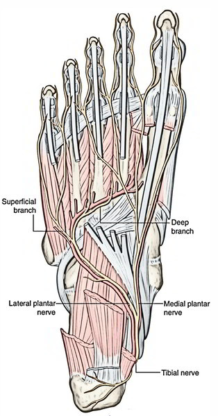
Medial Plantar Nerve
The medial plantar nerve is the major sensory nerve in the sole of the foot. It innervates skin on most of the anterior two-thirds of the sole and adjacent surfaces of the medial three and one-half toes, which includes the great toe. In addition to this large area of plantar skin, the nerve also innervates four intrinsic muscles-the abductor hallucis, flexor digitorum brevis, flexor hallucis brevis, and first lumbrical.
The medial plantar nerve passes into the sole of the foot deep to the abductor hallucis muscle and forward in the groove between the abductor hallucis and flexor digitorum brevis, supplying branches to both these muscles.
Lateral Plantar Nerve
The lateral plantar nerve is an important motor nerve in the foot because it innervates all intrinsic muscles in the sole, except for the muscles supplied by the medial plantar nerve (the abductor hallucis, flexor digitorum brevis, flexor hallucis brevis, and first lumbrical). It also innervates a strip of skin on the lateral side of the anterior two-thirds of the sole and the adjacent plantar surfaces of the lateral one and one-half digits.
The lateral plantar nerve enters the sole of the foot by passing deep to the proximal attachment of the abductor hallucis muscle. It continues laterally and anteriorly across the sole between the flexor digitorum brevis and quadratus plantae muscles, supplying branches to both these muscles, and then divides near the head of metatarsal V into deep and superfcial branches.
The superfcial branch of the lateral plantar nerve gives rise to a proper plantar digital nerve, which supplies skin on the lateral side of the little toe, and to a common plantar digital nerve, which divides to supply proper plantar digital nerves to skin on the adjacent sides of toes IV and V The proper plantar digital nerve to the lateral side of the little toe also innervates the flexor digiti minimi brevis and the dorsal and plantar interossei muscles between metatarsals IV and V.
The deep branch of the lateral plantar nerve is motor and accompanies the lateral plantar artery deep to the long flexor tendons and the adductor hallucis muscle. It supplies branches to the second to fourth lumbrical muscles, the adductor hallucis muscle, and all interossei except those between metatarsals IV and V which are innervated by the superfcial branch.
Motor Innervation of The Medial And Lateral Plantar Nerves
| Medial plantar nerve | Lateral plantar nerve |
|---|---|
| •Four muscles | • Flexor digitorum accessorius |
| • Abductor hallucis | • Abductor digiti minimi |
| • Flexor digitorum brevis | • Abductor hallucis |
| • Flexor hallucis brevis | • Flexor digiti minimi brevis |
| • First lumbrical | • All interossei |
| • Second, third, and fourth lumbricals |
Distribution of Pre-Axial And Post-Axial Nerves In The Palm And Sole
| Pre-axial nerves | Post-axial nerves | ||
|---|---|---|---|
| Median nerve | Medial plantar nerve | Ulnar nerve | Lateral plantar nerve |
| Abductor pollicis | Abductor hallucis | Abductor digiti minimi | Abductor digiti minimi |
| Flexor pollicis brevis | Flexor hallucis brevis | Flexor digiti minimi | Flexor digiti minimi brevis |
| Opponens pollicis | No corresponding muscle | Opponens digiti minimi | No corresponding muscle |
| Flexor digitorum superficialis | Flexor digitorum brevis | Abductor pollicis All interossei | Abductor hallucis All interossei Flexor digitorum accessorius |
| Lumbricals 1 and 2 | Lumbrical 1 | Lumbricals 3 and 4 | Lumbricals 2, 3, and 4 |
| 3Vi Digits | 3Vi Digits | P/2 Digit | P/2 Digit |
Plantar Arteries
There are 2 plantar arteries– medial and lateral.
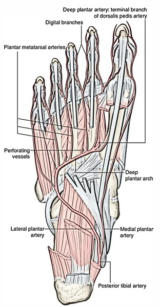
Medial Plantar Artery
The medial plantar artery is the smaller terminal branch of the posterior tibial artery. It originates below the flexor retinaculum and appears in the sole deep to abductor hallucis escorted by the medial plantar nerve on its lateral side.
Lateral Plantar Artery
The lateral plantar artery is the bigger terminal branch of the posterior tibial artery. It originates below the flexor retinaculum and runs forwards in the direction of the base of the fifth metatarsal. The lateral plantar nerve is located on its medial side. At the base of the fifth metatarsal bone, it curves medially with concavity facing proximally and runs in the direction of the very first inter-digital space to join the dorsalis pedis artery and so creates the plantar arch.
Test Your Knowledge
Sole of the foot

 (60 votes, average: 4.62 out of 5)