Boundaries
- Anterior: Deep fascia of the leg.
- Medial: Lateral surface of the shaft of the tibia.
- Lateral: Anterior intermuscular septum.
- Posterior: Interosseous membrane.
Contents
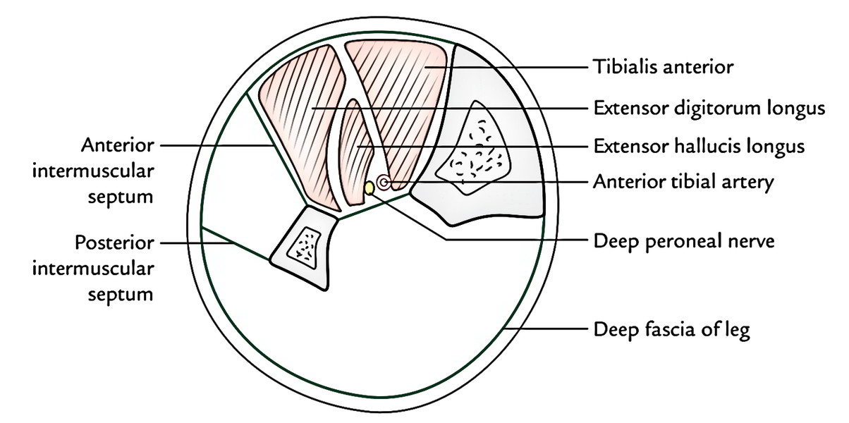
- Muscles: 4 muscles (tibialis anterior, extensor hallucis longus, extensor digitorum longus, and peroneus tertius).
- Artery: Anterior tibial artery.
- Nerve: Deep peroneal nerve (anterior tibial nerve).
Muscles of The Anterior Compartment of The Leg
With the exception of the Tibialis anterior which appears from tibia, all the muscles of the anterior compartment of the leg originate from the Fibula. All are supplied by the deep peroneal nerve and dorsiflex.
Origin, Insertion, and Actions of The Muscles of The Anterior Compartment of The Leg
| Muscle | Origin from | Insertion into | Actions |
|---|---|---|---|
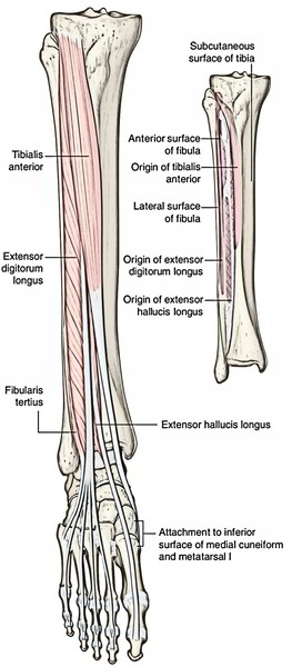
| Tibialis anterior (it has spindle-shaped multipennate belly) | 1. Upper 2/3rd of the lateral surface of the tibia 2. Adjacent part of the interosseous membrane 3. Its own covering deep fascia 3. Distal part of the lateral condyle of the tibia | 1. Medial surface of the medial cuneiform 2. Adjoining medial surface of the base of the 1st metatarsal | 1. Dorsiflexion of the ankle 2. Inversion of the foot 3. Maintenance of the medial longitudinal arch of the foot |
| Extensor hallucis longus | 1. Middle 2/4th (posterior part) of the medial surface of the shaft of fibula 2. Adjacent part of the interosseous membrane | Base (dorsal surface) of the distal phalanx of the big toe | 1. Extension of the phalanges of the big toe 2. Dorsiflexion of the foot |
| Extensor digitorum longus | Whole of the upper 1/4th and medial part of the middle 2/4th of the anterior surface of the fibula | Middle and distal phalanges of the lateral four toes by four tendons | 1. Dorsiflexion of the foot 2. Extension of MP*, PIP**, and DIP*** joints of the lateral four toes |
| Peroneus tertius | 1. Lower 1/4th of the anterior surface of the fibula 2. Adjacent part of the interosseous membrane | Dorsal surface (medial part) of the base of the 5th metatarsal bone | 1. Dorsiflexion of the foot 2. Eversion of the foot |
* MP: metatarsophalangeal, ** PIP = proximal interphalangeal, *** DIP = distal interphalangeal.
Tibialis Anterior
It’s a spindle-shaped multipennate muscle. It’s the most medial and superficial dorsiflexor of the foot, which is located against the lateral surface of the tibia.
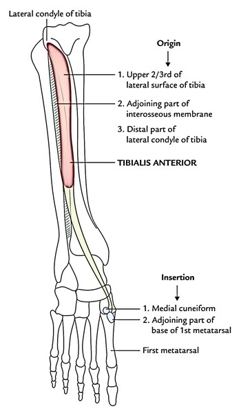
Origin
It appears from:
- Upper 2/3rd of the lateral surface of the tibia and adjoining part of the lateral condyle of the tibia.
- Adjoining part of the interosseous membrane.
- Overlying deep fascia of the leg.
Insertion
The muscle fibres converge below to create a tendon that is related to the lower 1/3rd of the lateral surface of the tibia. It pierces the medial part of superior extensor retinaculum and the upper group of inferior extensor retinaculum. Now it enters medially underneath the inferior group of inferior extensor retinaculum to be added on to the inferomedial side of the base of the very first metatarsal bone and adjacent part of the medial cuneiform.
Nerve Supply
By the deep peroneal nerve.
Actions
- It’s the leader dorsiflexor of the foot in the ankle joint.
- It keeps the medial longitudinal arch.
- It functions as an inverter of the foot at the midtarsal and subtalar joints.
Clinical testing
To analyze tibialis anterior medically, request the patient to stand on heels or dorsiflex the foot against resistance. If normal, its tendon can be viewed and palpated.
Clinical significance
- Anterior tibial compartment syndrome/shin splints (Fresher’s syndrome): It happens because of overexertion of the muscles of the anterior compartment particularly when the untrained men who lead a sedentary life are requested to walk or run for long distances. Shin splints also appear in trained runners who don’t warm up.
- Muscles of the compartment swell inside a tight compartment because of unexpected overuse that might impede venous return resulting in accumulation of more fluid in the compartment with unyielding walls. The pressure tends to compress the anterior tibial artery, and reducing the blood supply to the muscles, resulting in ischemia and pain. It’s often viewed in freshers (example, recently accepted medical students/ recently recruited military employees) who are made to run excessively. Therefore, this condition is also called military fresher’s syndrome.
Anterior Tibial Artery
The anterior tibial artery is the key artery of the anterior compartment of the leg. It corresponds to the posterior interosseous artery of the forearm. The blood supply to the anterior compartment of the leg is bolstered by the perforating branch of peroneal artery. Hence, the size of peroneal artery is inversely proportional to that of the anterior tibial artery.
The anterior tibial artery is escorted by 2 venae comitantes.
The anterior tibial artery descends through the anterior compartment on the interosseous membrane. In the distal leg, it lies between the tendons of the tibialis anterior and extensor hallucis longus muscles. It leaves the leg by passing anterior to the distal end of the tibia and ankle joint and continues onto the dorsal aspect of the foot as the dorsalis pedis artery.
In the proximal leg, the anterior tibial artery has a recurrent branch, which connects with the anastomotic network of vessels around the knee joint.
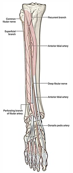
Deep Peroneal Nerve (Anterior Tibial Nerve)
It’s the nerve of the anterior compartment of the leg and dorsum of the foot. It corresponds to the posterior interosseous nerve of the forearm.
The nerve associated with the anterior compartment of the leg is the deep fibular nerve. This nerve originates in the lateral compartment of the leg as one of the two divisions of the common fibular nerve.
The deep fbular nerve passes anteromedially through the intermuscular septum that separates the lateral from the anterior compartments of the leg and then passes deep to the extensor digitorum longus. It reaches the anterio interosseous membrane where it meets and descends with the anterior tibial artery.
The deep fibular nerve innervates all muscles in the anterior compartment then continues into the dorsal aspect of the foot where it innervates the extensor digitorum brevis, contributes to the innervation of the first two dorsal interossei muscles, and supplies the skin between the great and second toes.

 (53 votes, average: 4.60 out of 5)