The vertebral column can be studied easily by comparing and contrasting the regions, numbers and the types of vertebrae namely cervical, thoracic, lumbar and sacral vertebrae within the vertebral column.
The vertebral column is comprised of the cervical, thoracic, lumbar, sacral and coccygeal vertebrae. The cervical vertebra can be found starting from the neck and to find the cervical vertebra. The vertebral column (spine or backbone) extends from the skull to the pelvis and forms a somewhat flexible but sturdy longitudinal support for the trunk. It is formed of 24 slightly movable vertebrae, the sacrum, and the coccyx. The vertebrae are separated from each other by intervertebral discs that serve as shock absorbers and allow bending of the spinal column. Four distinct curvatures can be seen on the lateral view of the vertebral column. From superior to inferior they are the cervical, thoracic, lumbar, and sacral curvatures. These curvatures provide flexibility and cushion, and allow the vertebral column to bear body weight more efficiently.
Structure of A Vertebra
Vertebrae are divided into three groups: cervical, thoracic, and lumbar vertebrae. Although each type has a distinctive anatomy, they have many features in common.
The anterior, drum-shaped mass is the body, which serves as the major load-bearing portion of a vertebra. A bony vertebral arch surrounds the large vertebral foramen through which the spinal cord and nerve roots pass. A spinous process
projects posteriorly and transverse processes project laterally from each vertebral arch.
A pair of superior articular processes
projects superiorly and a pair of inferior articular processes
projects inferiorly from the vertebral arch. The articular facet
(fa^set) of each superior articular process articulates with the articular fact of the inferior articular process of the adjacent vertebra superior to it. When joined by ligaments, the vertebrae form the vertebral canal
that protects the spinal cord.
Small intervertebral foramina
occur between adjacent vertebrae. They serve as lateral passageways for spinal nerves that exit the spinal cord.
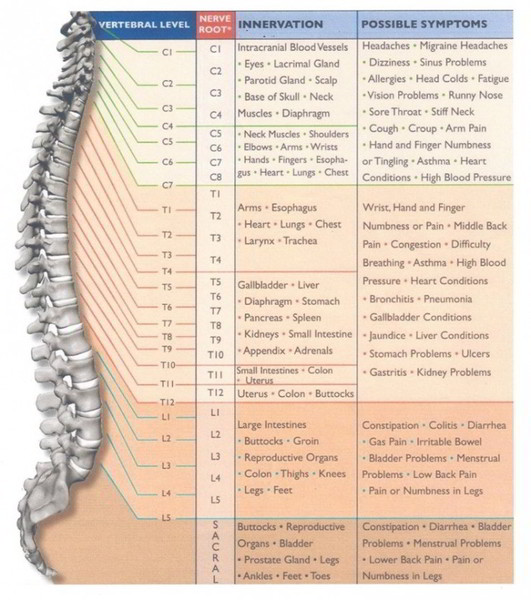
Cervical Vertebrae
The cervical vertebrae can be found at the base of the skull, the occipital bone and the first rib and the seventh cervical vertebra between the two landmarks (base of the skull and rib one) where the first and third thoracic vertebra is located. So C 1 is the first vertebra and this vertebrae ends all the way down at c7.
The first seven vertebrae support the neck. They are unique in having a transverse foramen in each transverse process. It serves as a passageway for the vertebral arteries and veins, blood vessels involved in blood flow to and from the brain.
The first two cervical vertebrae are distinctly different from the rest. The first vertebra (C1), or atlas, whose superior articular facets articulate with the occipital condyles, supports the head. The second vertebra (C2), which is called the axis, has a prominent dens
that projects superiorly from the vertebral body, providing a pivot point for the atlas. When the head is turned, the atlas rotates on the axis.
Thoracic Vertebrae
The thoracic vertebrae as the name suggests are located in the thoracic region. They can be found quite easily because there are 12 pairs of ribs that come off of each of these vertebrae. Counting from rib one to rib twelve gives us the first and last thoracic vertebrae which means there’s 12 of them and are numbered sequentially t1 to t12.
The thoracic vertebrae (T1-T12) are larger than the cervical vertebrae, and their spinous processes are longer and slope inferiorly. The ribs articulate with costal facets on the transverse processes and bodies of thoracic vertebrae.
Lumbar Vertebrae
The lumbar vertebrae are located in the lower back and these are found at the first vertebra which is below the the first rib and then all the way down to the last articulating rib after which the sacrum is fused below. To make it easier to find it, the vertebra below the first rib is where the lumbar vertebrae starts which are numbered as l1, l2, l3, l4 and l5 vertebra.
The five lumbar vertebrae (L1-L5) have heavy, thick bodies to support the greater stress and weight that is placed on this region of the vertebral column. The spinous processes are blunt and provide a large surface area for the attachment of heavy back muscles.
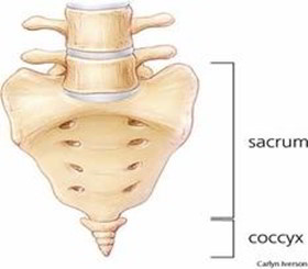
Sacrum
The sacrum is composed of five fused vertebrae but even though they’re fused, there’s still a segmental organization numbered as the S1, S2, S3, S4 and S5 sacral levels and this is significant because they still have the spinal nerves that exit below their associated intervertebral frame.
It articulates with the fifth lumbar vertebra and forms the posterior wall of the pelvis. The spinous processes of the fused vertebrae form the median sacral crest on the posterior midline. On either side of the median sacral crest are the posterior sacral foramina, passageways for blood vessels and nerves. Anterior sacral foramina on the anterior surface serve a similar function. The sacral canal is a continuation of the vertebral canal that carries spinal nerve roots to the sacral foramina and the sacral hiatus, an inferior opening proximal to the coccyx.
Coccyx
There are five fused vertebrae that make up the tailbone, the coccygeal vertebrae. This completes the vertebral column and in each of the regions, the vertebral column consists of some curvature which are referred to as primary and secondary curvatures.
Primary and Secondary Curvatures
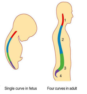
First is the primary curvature. In the picture of fetus above, it can be noticed that the fetus is in the fetal position and the vertebra makes this primary curve which is a C shaped curve and it’s primary because it’s the first curve is in the verbal column. The curve is retained in the thoracic region of the vertebral column and so the curve in the thoracic vertebra is called the primary curvature
In adults, the cervical and lumbar regions, there are secondary curves because they come after the primary curve in the cervical and lumbar region and these are also called lordotic curves or lordotic curvatures. And when there is an over curvature in lumbar region, it is referred to as lordosis.

 (58 votes, average: 4.71 out of 5)