Vision is one of the most important senses supplying information to the brain. The sensory receptors for light stimuli are located within the eyes (or eyeballs), the organs of vision. The eyes are located within the orbits, where they are protected by seven skull bones Connective tissues provide support and protective cushioning for the eyes.
Eyelids, Eyelashes, and Eyebrows
The exposed anterior surface of the eye is protected by the eyelids. Blinking spreads tears and mucus over the anterior eye surface to keep it moist. The internal surface of each eyelid is lined with a mucous membrane called the conjunctiva (kon-junk-ti ‘-vah), which continues across the anterior surface of the eye. Only its transparent superficial epithelium covers the cornea. Mucus from the conjunctiva helps to lubricate the eye and keep it moist. The conjunctiva also contains many blood vessels and nociceptors.
Eyelashes help to keep airborne particles from reaching the eye surface and provide some protection from excessive light. Eyebrows, located on the brow ridges, also shield the eyes from overhead light and divert sweat from the eyes. Observe the accessory structures in figure 9.12.
Lacrimal Apparatus
The lacrimal (lak’-ri-mal) apparatus, shown in figure 9.13. is involved in the production and removal of tears. Tears are secreted continuously by the lacrimal gland, which is located in the superior, lateral part of each orbit. Tears are carried to the surface of the eye by a series of tiny excretory ducts. The tears flow inferiorly and medially across the eye surface as they are spread over the eye surface by blinking. Once collected at the medial corner of the eye by the lacrimal canaliculi, tears flow into the lacrimal sac, and flow on through the nasolacrimal duct into the nasal cavity.
Tears perform an important function in keeping the anterior surface of the eye moist and in washing away foreign particles. An antibacterial enzyme (lysozyme) in tears helps to reduce the chance of eye infections.
Extrinsic Muscles
Movement of the eyes must be precise and in unison to enable good vision. Each eye is moved by six extrinsic muscles of the eyeball that originate from the posterior of the orbit and insert on the surface of the eye. Four muscles exert a direct pull on the eye, but two muscles pass through cartilaginous loops, enabling them to exert an oblique pull on the eyeball. Although each muscle has its own action, these muscles function as a coordinated group to enable eye movements. The locations and functions of these muscles are shown in figure 9.14.
Structure of the Eye
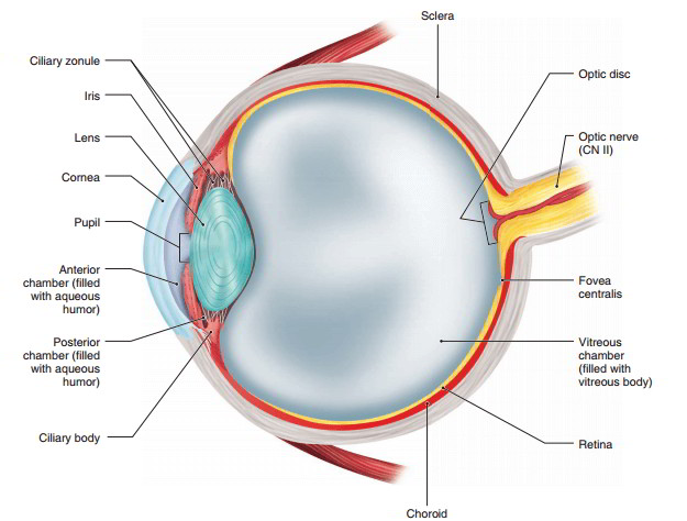
The eye is a hollow, spherical organ about 2.5 cm (1 in) in diameter. It has a wall composed of three layers and internal spaces filled with fluids that support the walls and maintain the shape of the eye. The major parts of the eye are shown in figure 9.15.
External Layer The external layer of the eye consists of two parts: the sclera and the cornea. The sclera (skle ‘-rah) is the opaque, white portion of the eye that forms most of the external layer. The sclera is a tough, fibrous layer that provides protection for the delicate internal portions of the eye and for the optic nerve (CN II), which emerges from the posterior portion of the eye. The anterior portion of the sclera is covered by the conjunctiva. The cornea
(kor’-ne-ah) is the anterior clear window of the eye. It has a greater convex curvature than the rest of the eyeball so that it can bend light rays as they pass through it. It lacks blood vessels and nerves that would block light rays from entering the eye.
Middle Layer The middle layer includes the choroid, ciliary body, and iris. The choroid (ko’-roid), which is found in all but the anteriormost portion of the layer, contains blood vessels that nourish the eye and large amounts of melanin. The absorption of light by melanin prevents back-scattering of light, which would impair vision. The ciliary (sil’-e-ar-e) body contains the ciliary muscles and forms a ring around the l ens just anterior to the choroid. The ciliary zonule contains fibrous strands that extend from the ciliary body to the lens and hold the lens in place. Contraction and relaxation of ciliary muscles change the shape of the lens.
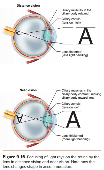
Although entering light rays are bent by the cornea, it is the lens that focuses light rays precisely on the retina. The transparent, somewhat elastic lens is composed of protein fibers and lacks blood vessels and nerves that would block the passage of light rays. Contraction and relaxation of the ciliary muscles change the shape of the lens in a process called accommodation (figure 9.16). Contraction of the ciliary muscles relaxes the fibrous strands in the ciliary zonule and allows the lens to become more spherical in shape. The relaxation of the ciliary muscles increases tension on the fibrous strands of the ciliary zonule and causes the lens to take on a more flattened shape. In this way, the shape of the lens is adjusted for distant, intermediate, and near vision so that the image is focused precisely on the retina.
The colored portion of the eye is the iris, a thin disc of connective tissue and smooth muscle that extends from the ciliary body anterior to the lens. The iris controls the amount of light entering the eye by controlling the size of the pupil. The pupil is the opening in the center of the iris through which light passes to the lens. Its size is constantly adjusted by the iris as lighting conditions change. The pupil is constricted in bright light and is dilated in dim light.
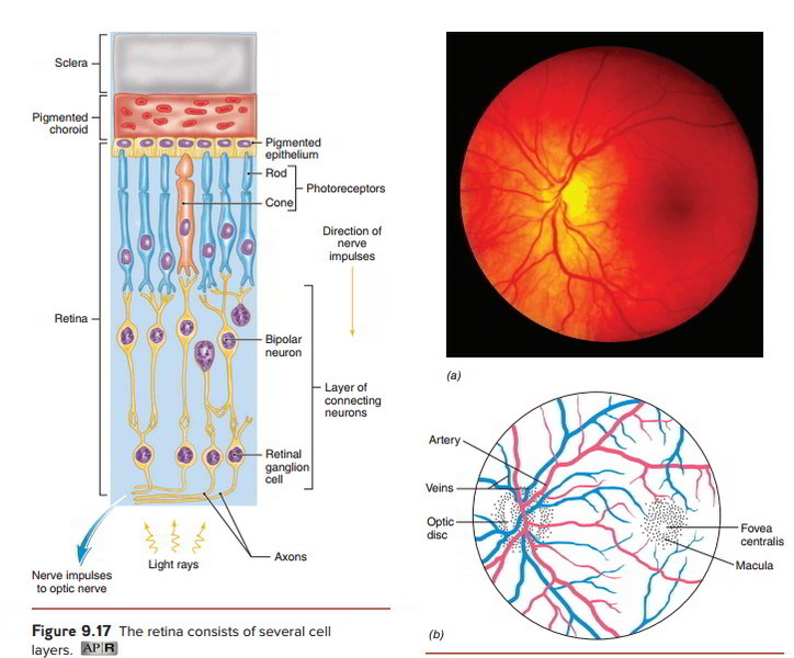
Internal Layer The filmlike retina (ret’-i-nah) lines the internal surface of the eye posterior to the ciliary body. The retina contains two types of photoreceptor cells: rods and cones. Rod and cone anatomy can be seen in figure 9.17 . The thin, elongate rods are photoreceptors for black and white vision because they are sensitive only to the presence of light. The shorter and thicker cones are photoreceptors for color vision. Because cones require bright light to function, only rods allow us to see in dim light.
The macula (mak’-u-lah) is a yellowish disc on the retina directly posterior to the lens. In the center of the macula, there is a small depression called the fovea centralis (fo’-ve-ah sen-trah’-lis). The fovea centralis contains densely packed cones, making it the area for the sharpest color vision. The density of the cones decreases with increased distance from the fovea. Rods, which are absent from the fovea, increase in density with increased distance from the fovea. Therefore, dim-light vision is best at the edge of the visual field (figure 9.18; see figure 9.15). It is important to remember that the macula on the retina is structurally and functionally different from the maculae within the internal ear.
The retina contains neurons in addition to rods and cones (see figure 9.17) . Nerve impulses formed by rods and cones are transmitted to retinal ganglion cells, whose axons converge at the optic disc to form the optic nerve. The optic disc is located medial to the fovea. Because the optic disc lacks photoreceptors, it is also known as the “blind spot.” However, we usually do not notice a blind spot in our field of vision because the visual fields of our eyes overlap.
An artery enters the eye and a vein exits the eye via the optic disc. These blood vessels are continuous with capillaries that nourish the internal tissues of the eye and are the only blood vessels in the body that can be viewed directly. A special instrument called an ophthalmoscope (of-thal’-mo-skop) is used to look through the lens and observe these vessels. Figure 9.18 shows the appearance of blood vessels and the retina as viewed with an ophthalmoscope.
Internal Cavities The space between the cornea and the iris is known as the anterior chamber, which is filled with a watery fluid called aqueous (a’-kwe-us) humor. The small posterior chamber, located between the iris and lens, is also filled with aqueous humor. The aqueous humor is filtered out of capillaries in t he ciliary body, flows through the posterior chamber into the anterior
chamber, and is reabsorbed into blood vessels located at the junction of the sclera and cornea. Aqueous humor is largely responsible for the internal pressure within the eye and the normal shape of the cornea. The aqueous humor also provides nourishment to the cornea and lens. Normally, it is secreted and absorbed at the same rate so that intraocular pressure is maintained at a constant level.
The large vitreous chamber is located posterior to the lens. It is filled with a clear, gel-like substance called the vitreous (vit’-re-us) body. The vitreous body, which forms during embryonic development, is not reabsorbed or regenerated. The vitreous body presses the retina firmly against the wall of the eye and helps to maintain the shape of the eye.
Physiology of Vision
Light rays coming to the eye must be precisely bent so that they are focused on the retina. This bending of the light rays is called refraction (re-frak’-shun) and it is produced by the cornea and lens. The convex surface of the cornea produces the greatest refraction of light rays, while further bending (accommodation) by the lens provides a “fine adjustment” so that the image is focused precisely on the retina.
The optics of the eye cause the image to be inverted on the retina, as shown in figure 9.16 . However, the visual areas of the cerebral cortex correct for this inversion so that objects are seen in their correct orientation. When images are incorrectly focused on the retina, poor vision results. Figure 9.19 shows common optical disorders and how they may be corrected with glasses, contact lenses, or Lasik surgery.
When light rays strike the retina, the light stimuli must be converted into nerve impulses that are sent to the brain. Both rods and cones contain light-sensitive pigments that break down into simpler substances when light is absorbed. The breakdown of these pigments results in the formation of nerve impulses.
Rods contain a light-sensitive pigment called rhodopsin that breaks down into opsin, a protein, and retinal, which is derived from vitamin A. This breakdown triggers the formation of nerve impulses that are carried via the optic nerve to the brain. Rhodopsin is resynthesized from opsin and retinal to prepare the rods for receiving subsequent stimuli. A deficiency of vitamin A mayresult in an insufficient amount of rhodopsin in the rods, which, in turn, may lead to night blindness, the inability to see in dim light.
Although the light-sensitive pigments are different in cones, they function in a similar way to rhodopsin. There are three different types of cones, and each has a pigment that responds best to a different color (wavelength) of light. One type responds best to red light, another type responds best to green light, and the third type responds to blue light. The perceived color of objects results from the combination of the cones that are stimulated and the interpretation of the nerve impulses that they form by the cerebral cortex.
Nerve Pathway Nerve impulses formed by the photoreceptors are transmitted via axons of the optic nerve to the brain. The optic nerves merge just anterior to the pituitary gland to form an X-shaped pattern called the optic chiasma (ki ‘-as-mah) (figure 9.20). Within the optic chiasma, the axons from the medial half of the retina in each eye cross over to the opposite side. Thus, the medial axons of the left eye and the lateral axons of the right eye form the right optic tract leaving the optic chiasma. Similarly, the medial axons of the right eye and the lateral axons of the left eye form the left optic tract leaving the optic chiasma. The axons of the optic tracts enter the thalamus, where they synapse with neurons that carry the nerve impulses on to the visual areas of the occipital lobes.
The crossing of the medial axons results in each visual area receiving images of the entire object but from slightly different views, which create stereoscopic (threedimensional) vision. Depth perception is a result of stereoscopic vision.
The mechanism of vision may be summarized as follows (figure 9.20):
- Light rays are bent as they pass through the cornea.
- The iris controls the amount of light passing through the pupil.
- The ciliary body adjusts the shape of the lens to focus the light rays (image) on the retina.
- Light absorbed by the rods and cones causes the formation of nerve impulses.
- The nerve impulses are transmitted to neurons whose axons converge at the optic disc to form the optic nerve.
-
The medial axons of the optic nerves cross over at the optic chiasma and merge with the lateral axons on that side to form the optic tracts, which continue to the thalamus.
The nerve impulses are then carried to the vision areas in the occipital lobes of the cerebrum, where they are interpreted as visual images.
Disorders of The Eye
Astigmatism (a-stig’-mah-tizm) is the unequal focusing of light rays on the retina, which causes part of an image to appear blurred. It results from an unequal curvature of the cornea or lens.
Blindness is partial loss or lack of vision. It may be caused by a number of disorders such as cataract, glaucoma, and detachment or deterioration of the retina. It may also result from damage to the optic nerves or the visual centers in the occipital lobes of the cerebrum.
Cataract is cloudiness or opacity of the lens, which impairs or prevents vision. It is common in older people and is the leading cause of blindness. Surgical removal of the clouded lens and implantation of a plastic lens usually restores good vision.
Color blindness is the inability to perceive certain colors or, more rarely, all colors. Red-green color blindness, the most common type, is characterized by difficulty distinguishing reds and greens due to the absence of either red or green cones. Color blindness is inherited, and it occurs more often in males than in females because it is a sex-linked hereditary trait.
Conjunctivitis (con-junk-ti-vi ‘-tis) is inflammation of the conjunctiva. It may be caused by allergic reactions, physical or chemical causes, or infections. Inflammation that results from a bacterial or viral infection is commonly called pink eye. Such bacterial infections are highly contagious.
Farsightedness (hyperopia) is blurred vision caused by light rays being incorrectly focused posterior to the retina. Its causes include lens abnormalities and the eye being shorter than normal.
Nearsightedness (myopia) is blurred vision caused by light rays being incorrectly focused anterior to the Retina. Its causes include lens abnormalities and the eye being longer than normal.
Presbyopia (prez-be-o’pe-ah) is the diminished ability of the lens to accommodate for near vision due to a decrease in its elasticity. It is a natural result of aging. At age 20, an object can be clearly observed about 10 cm (4 in) from the eye. At age 60, an object must be about 75 cm (30 in) from the eye to be clearly observed.
Retinoblastoma (ret-i-no-blas-to’-mah) is a cancer of immature retinal cells. It constitutes about 2% of the cancers in children.
Strabismus (strah-biz’-mus) is a disorder of the extrinsic eye muscles in which the eyes are not directed toward the same object simultaneously. Treatment may include eye exercises, corrective lenses, or corrective surgery.

 (62 votes, average: 4.82 out of 5)