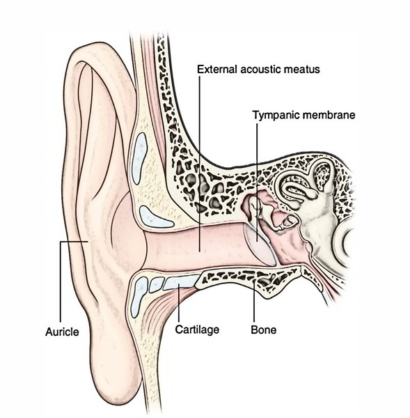
The external ear is composed of (a) pinna or auricle and (b) external auditory meatus that are concerned with collection and transmission of sound waves to the tympanic membrane, respectively.
Auricle/Pinna
The auricle is located on the side of the head and is trumpet-like undulating projection. With the exception of its lobule the whole pinna is composed of a single bit of crumpled yellow elastic cartilage covered with skin. The lobule of pinna is made of fibrofatty tissue covered with skin. The auricular cartilage stays constant with the cartilage of the external auditory meatus.
Features
The auricle presents 2 surfaces: medial and lateral.
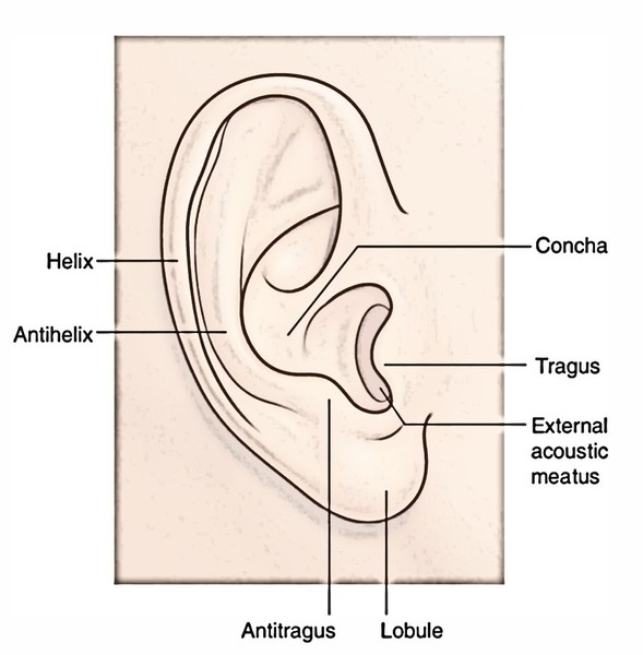
The lateral surface of auricle exhibits following natural elevations and depressions:
- Concha, a large depression that leads into the external auditory meatus. It’s guarded in front by a triangular flap of cartilage, the tragus.
- Helix creates a notable peripheral rim of the pinna. It includes 2 limbs-anterior and posterior. An anterior limb ends as crus of helix, which breaks up the concha into smaller upper and bigger lower parts. The posterior limb finishes below as flabby ear lobe and its upper end occasionally presents a small elevation referred to as Darwin’s tubercle. It’s likely erroneously thought to represent the vestige of the pointed part of the quadruped ear.
- Antihelix is just another notable ridge present in front and parallel to the posterior part of helix, partially encircling the concha. Its upper end splits into 2 crura enclosing a triangular depression named triangular fossa. The narrow gutter between the helix and antihelix is named scaphoid fossa.
Tragus is a small triangular flap before concha.
Antitragus is a small elevation opposite to tragus from which it’s divided by an intertragic notch.
Cymbaconchae is a small area of concha above the crus of helix. Medically it’s significant as it corresponds to the suprameatal triangle (McEwen’s triangle).
Lobule of the ear hangs below the antitragus as a large skin covered flap of fibrofatty tissue.
Key Points
- There’s no cartilage between tragus and crus and the gap between the 2 is termed incisuraterminalis.
- The thick hair on pinna especially on tragus in male represents Y-linked inheritance.
- The pinna collects and directs the sound waves to the external auditory meatus.
- The medial/cranial surface of (pinna) presents the followingfeatures:
- Eminentia concha, which corresponds to the depression of the concha.
- Eminentiatriangularis, which corresponds to the triangular fossa between the crura of the antihelix.
Clinical Significance
Pinna is a source of several graft materials for the surgeons.
The lobule of ear is often pierced for wearing earrings.
Muscles
For surgery of external auditory meatus, the incision is created in the region of incisuraterminalis as it WOn’t cut via the cartilage.
The extrinsic muscles pass from scalp or skull to the auricle. They’re as follows:
- Auricularis anterior.
- Auricularis superior.
- Auricularis posterior.
The anterior and superior muscles originate from epicranialaponeurosis and are added into the upper part of the helix and upper part of the cranial outermost layer of the auricle, respectively. The auricularis posterior appears from the mastoid process and gets fit into eminentia concha.
The intrinsic muscles are small muscular skids, which pass between the cartilaginous parts of the auricle.
Activities
The extrinsic muscles may play a part in positioning of the auricle to catch the sound, while intrinsic muscles may alter the shape of the auricle. Such movements are seldom viewed in human beings. Nevertheless in creatures they change the shape of the pinna.
Skin
The skin covering the auricle is closely adherent to the underlying cartilage and fibrofatty tissue. Occasionally rough hair projects out of the tragus, antitragus, heartbreaking notch and helix in aged men. The hairy pinna is an expression of Y-linked genes.
Arterial Supply
The cranial surface and posterior part of the lateral surface is supplied by the posterior auricular branch of the external carotid artery.
The anterior part of the lateral surface is supplied by the superficial temporal artery.
Few branches of occipital artery furnish the upper part of the cranial surface.
Venous Drainage
The veins accompany the arteries and drain into superficial temporal and external jugular veins.
Lymphatic Drainage
The lymph from auricle drains into:
- Preauricular (parotid) lymph nodes.
- Mastoid lymph nodes.
- Upper group of deep cervical lymph nodes.
Nerve Supply
- Motor Supply: All the extrinsic and intrinsic muscles of the auricle are supplied by the facial nerve. The auricularis anterior and auricularis superior are supplied by the temporal branch of the facial nerve, while auricularis.
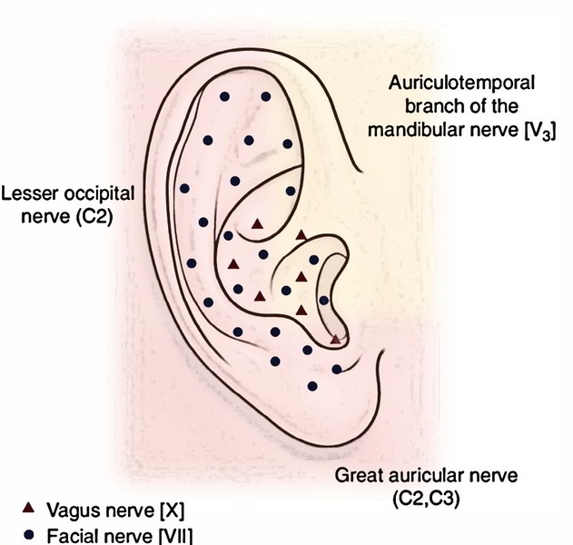
- Lateral (facial) surface
- Lower one-third, by great auricular nerve (C2, C3).
- Upper two-third, by auriculotemporal nerve [(a branch of mandibular division of the trigeminal nerve (CNV)]
- Concha, by auricular branch of the vagus (Alderman’s nerve) nerve (CNX).
- Medial (cranial) surface.
- Lower one-third, by great auricular nerve (C2, C3).
- Upper two-third, by lesser occipital nerve (C2).
- Eminentia conchae, by auricular branch of the vagus.
Clinical Significance
Engagement of pinna in herpes zoster of geniculate ganglion (Ramsay Hunt syndrome): Medically, it’s recognized that a couple fibres of the facial nerve accompany the auricular branch of vagus and supply the skin in the region of concha and eminentia conchae, as vesicles are found in these regions during engagement of the geniculate ganglion of the facial nerve by herpes zoster virus. The communication between the auricular branch of vagus and facial nerves happens inside the petrous temporal bone.
External Auditory Meatus
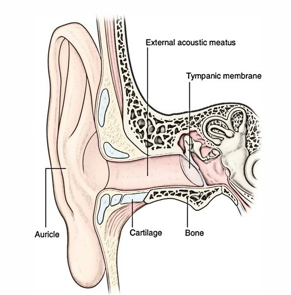
The external auditory meatus (syn. external acoustic meatus) stretches from the base of the concha to the tympanic membrane and measures about 24 millimeters along its posteriorwall. Notice that it’s not a straight tube but it’s a typical S-shaped course. Its outer part is pointed upwards, backwards and medially (UBM), on the other hand its inner part is pointed downwards, forwards and medially (DFM). For that reason, to examine the tympanic membrane the pinna needs to be pulled upwards, backwards and laterally, to bring the 2 parts in alignment.
Parts
The external auditory meatus is split into 2 parts: cartilaginous and bony.
The cartilaginous part creates the outer one-third (8 millimeters) of the meatus. The cartilage is the continuance of the cartilage of the auricle. The skin covering the cartilaginous part is thick and includes hair and ceruminous (pilosebaceous) glands, which secrete ear wax.
The bony part creates the inner two-third (16 millimeters) of the external auditory meatus. The skin lining the bony part of meatus is thin and constant with the cuticular layer of the tympanic membrane. It’s devoid of hair and ceruminous glands. About 4 mm lateral to the tympanic membrane (about 20 millimeters deep to concha), the bony meatus presents a narrowing termed isthmus. The foreign body lodged medial to isthmus gets influenced and are not easy to eliminate.
Occasionally the anterior wall of bony part presents a foramen (foramen of Huschke), allowing infection back and forth from parotid gland. This foramen is normally present in kids up to the age of FOUR years.
In the newborn, the bony canal isn’t developed and is represented by a tympanic ring of bone. Thus the external auditory meatus is shorter in children and consequently, deep insertion of ear speculum may damage the tympanic membrane.
Clinical Significance
Since the hairs are confined to the outer part of the meatus, the furuncles (infection of hair follicles) grow only in this part.
To examine external auditory meatus and tympanic membrane, the pinna is pulled upwards, backwards and laterally (vide supra) in adults, while in babies it’s pulled downwards and backwards. This is because in babies the bony part of external auditory meatus isn’t developed and tympanic membrane is pointed primarily downwards.
Arterial Supply
The external auditory meatus is supplied by these arteries:
- Deep auricular artery, a branch of first part of the maxillary artery.
- Anterior tympanic artery, a branch of first part of the maxillary artery.
Nerve Supply
Roof and anterior wall are supplied by the auriculo-temporal nerve.
Floor and posterior wall are supplied by the auricular branch of vagus (notice that it’s the only cutaneous branch of the vagus nerve).
Clinical Significance
The infection and boils of the external auditory meatus cause very little swelling but are really distressing since the skin liner is securely stuck to the underlying cartilage and bone.
Ear wax: It prevents the injury of the lining epithelium of the external auditory meatus from water and the damage of tympanic membrane by trapping the insects. The surplus of ear wax interfering with hearing is removed by syringing. The aggravation of auricular branch of vagus during syringing may reflexly generate constant cough referred to as ear cough, vomiting and sometimes even death because of sudden cardiac inhibition.
The Aldermen were the people in early Rome, who were quite fond of excessive eating and utilized to arouse their jaded hunger by dropping chilly water or spirit supporting the ear as this could reflexly arouse gastric peristalsis because of supply of the area by the vagus nerve which also supplies motor innervation to the GIT.
Development
The external auditory meatus grows as an ectodermal invagination of first pharyngeal cleft. It becomes filled up with ectodermal cells creating a solid mass termed meatalstopper that is canalized before arrival. The auricle grows from 6 mesodermal tubercles around the external opening of the very first pharyngeal cleft. The failure of canalization of meatal stopper leads to atresia of the external auditory meatus, while failure of fusion of tubercles will generate accessory auricles.
Tympanic Membrane
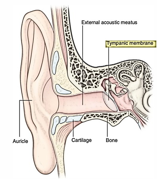
The tympanic membrane (or ear drum) is a thin (0.1 mm thick) semitransparent membrane, which creates the partition between external acoustic meatus and middle ear. It’s oval, measuring 9-10 millimeter in length and 8-9 mm in width. It’s set obliquely making an angle of about 55 ° with the floor of the external acoustic meatus. The tympanic membrane faces downwards, forwards and laterally as though to catch the sounds reflected from the earth. Therefore the anterior wall and the floor of external auditory meatus are longer in relation to the posterior wall and the roof.
Structure
The tympanic membrane is created of 3 layers from lateral to medial these are as follows:
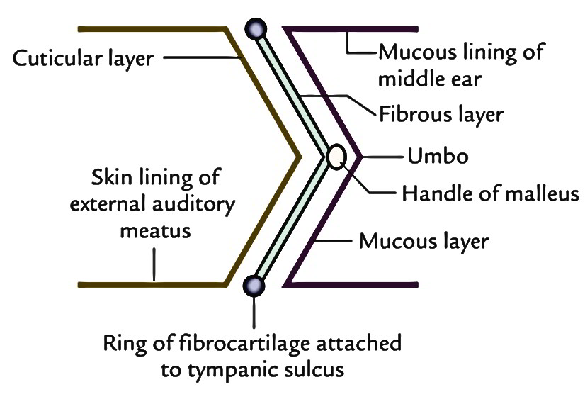
- Outer cuticular layer of stratified squamous epithelium that is constant with the skin lining the external auditory, meatus.
- Middle fibrous layer, which encloses the handle of the malleus. It includes outer radiating and inner circular fibres.
- Inner mucosal layer is lined by low columnar epithelium that is constant with the mucous lining of the middle, ear.
Parts
The tympanic membrane is split into 2 parts: pars tensa and pars flaccida.
Pars tensa creates majority of the tympanic membrane. Its periphery is thickened to create a fibrocartilaginous rim termed annulus tympanicus, which fits into the tympanic sulcus. The fibrocartilaginous rim presents a notch above. From the margins of the notch the anterior and posterior malleolar folds in mucous membrane of tympanic cavity pass to obtain connection to the lateral process of the malleus. The handle of the malleus is securely connected to the inner surface of the pars tensa. This part is left rough by the inward pull of the tensor tympani muscle, connected to the root of handle of the malleus and radial fibres.
Pars flaccida (Shrapnell’s membrane) is a small triangu-lar area above the lateral process of malleus between anterior and posterior malleolar folds (currently named malleal folds). This part is thin and lax because intermediate fibrous layer here is replaced by loose areolar tissue. It seems somewhat pinkish.
Surfaces
Lateral surface of the tympanic membrane is concave in the direction of the meatus and pointed downwards, forwards and laterally.
Medial surface is convex and bulges in the direction of the middle ear. The stage of maximum convexity is named umbo . When the tympanic membrane is illuminated forinspection, the concavity of the membrane creates a ‘cone of light radiating from the umbo over the anteroinferior quadrant. This surface gets the connection of malleus up to the centre of the membrane. Here the handle of the malleus is crossed medially by chorda tympani nerve, which runs forwards between the fibrous and mucosal layer in the junction of pars flaccida and pars tensa.
Arterial Supply
The outer surface is supplied by deep auricular artery, a branch from first part of maxillary artery.
The inner surface is supplied by (a) anterior tympanic artery, a branch from first part of maxillary artery and (b) posterior tympanic artery, a branch from stylomastoid artery originating from posterior auricular artery.
Venous Drainage
Veins from outer surface drain into external jugular vein.
Veins from inner surface drain into transverse sinus and pterygoid venous plexus.
Nerve Supply
Anterior half of the lateral surface is supplied by the auriculotemporal nerve (V3).
Posterior half of the lateral surface by the auricular branch of vagus (CNX).
Medial surface by tympanic branch of the glossopharyngeal (CNIX) via tympanic plexus.
Clinical Significance
Perforation of the tympanic membrane: It may result from an external injury or middle ear infection (otitis media).
Evaluation of tympanic membrane: Review of the tympanic membrane with an otoscope gives critical info regarding the state of the middle ear. The shade, curvature, presence of lesions and position of malleus are features of exceptional significance. When tympanic membrane is illuminated for evaluation, a cone of light is reflected in the anteroinferior quadrant of the membrane from umbo, the point of maximum concavity, which indicates the connection of the handle of the malleus. Since the membrane is see-through, these structures being located deep to it can be observed:
- Handle of Malleus, as a yellowish stripe going from umbo upwards and forwards.
- Sidelong process of malleus, as a white bulge in the upper part of the stripe of handle of malleus.
- Long processes of incus, as white stripes behind and parallel to the upper part of the handle of malleus.
A cone of light at 5 o’clock position in anteroinferior quadrant. Medically, the tympanic membrane is split into 4 quadrants by means of 2 imaginary lines going through the umbo. 1 is drawn along the handle of the malleus and the other at right angle to it via the umbo.
On illumination the normal membrane appears pearly gray. Occasionally an incision is supplied in the tympanic membrane (myringotomy) to drain the pus from the middle ear. The incision is generally supplied in the posteroinferior quadrant to prevent injury to the chorda tympani nerve, which crosses the inner aspect of the membrane in the upper part.
Development
The tympanic membrane grows from first pharyngeal membrane consisting, from superficial to deep, of 3 layers: ectoderm, mesoderm and endoderm.
Thus, the tympanic membrane also contains 3 layers from superficial to deep these are:
- Cuticular layer, originated from ectoderm.
- Intermediate layer, originated from mesoderm.
- Mucous layer, originated from endoderm.
The 3 layers of tympanic membrane are likened to the 3 layers of trilaminar embryonic disc.


 (71 votes, average: 3.73 out of 5)