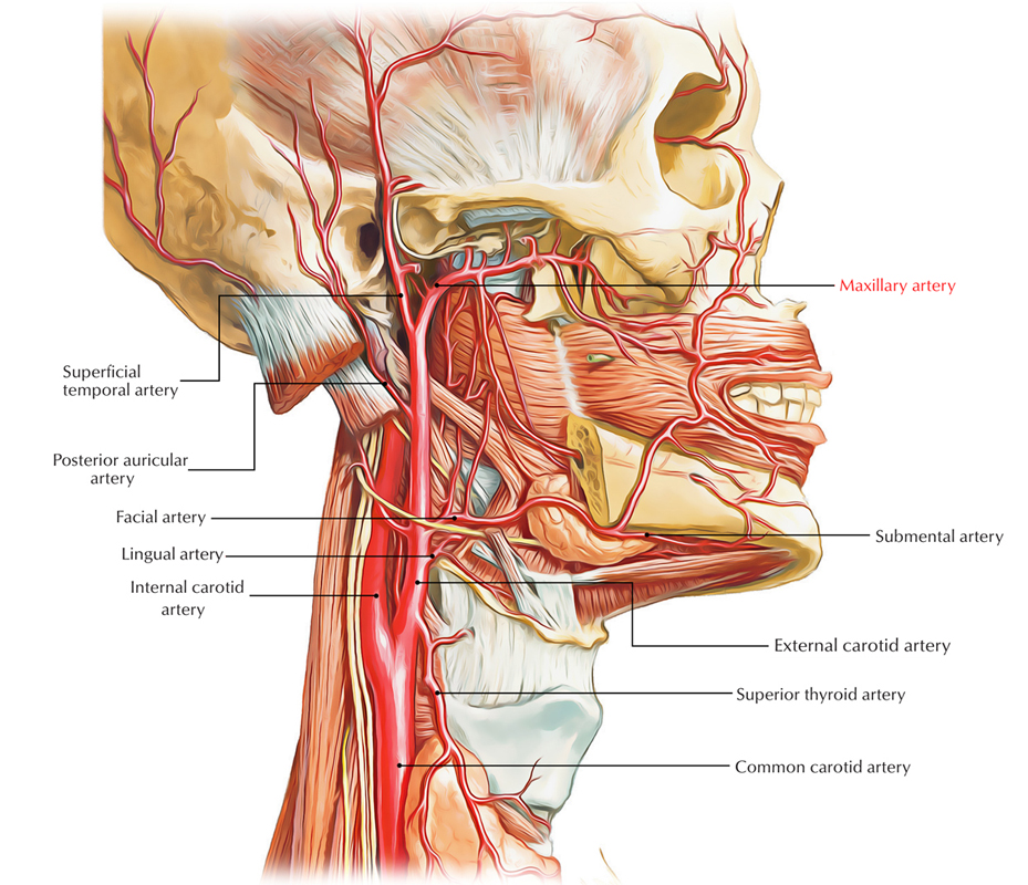
The maxillary artery is the bigger terminal branch of the external carotid artery.
It runs horizontally forward up to the lower border of lower head of lateral pterygoid and the point of origin is behind the neck of the mandible. Now it crosses the lower head of lateral pterygoid superficially (occasionally deep) after turning upwards and forwards. It enters the pterygopalatine fossa by going through pterygomaxillary fissure after coming between both heads of lateral pterygoid. Here it finishes by getting divided into its terminal branches.

Maxillary Artery
The maxillary artery has a wide land of distribution. It supplies:
- Upper and lower jaws,
- Muscles of temporal and infratemporal fossae,
- Nose and paranasal sinuses,
- Palate and roof of pharynx,
- External and middle ear,
- Pharyngotympanic tube and
- Dura mater.
The maxillary artery enters the temple fossa by passing forwards, between the neck of mandible and the sphenomandibular ligament.
Parts and Connections
The maxillary artery is split into 3 parts by the lower head of lateral pterygoid muscle. The parts are:
First part (mandibular part): From start (origin) to lower border of lateral pterygoid. It is located between the neck of the mandible laterally and sphenomandibular ligament medially. The auriculotemporal nerve is located above this part.
Second part (pterygoid part): From lower border to the upper border of the lower head of lateral pterygoid (i.e., second part is located on or deep to lower head of lateral pterygoid).
Third part (pterygopalatine part): From upper border of the lower head of lateral pterygoid to pterygopalatine fossa. In pterygopalatine fossa it is located in front of the pterygopalatine ganglion.
Key Points
- The Majority of the branches from the first and second parts of maxillary artery accompany the branches of the mandibular nerve.
- Branches from the third part of the maxillary artery accompany the branches of maxillary nerve and pterygopalatine ganglion.
- Branches from the second part of the maxillary artery are muscular only and supplies muscles of mastication.
- All the branches (first and third part) of the maxillary artery go through bony foramina and fissures with the exception of branches from its second part.
Branches of the Maxillary Artery

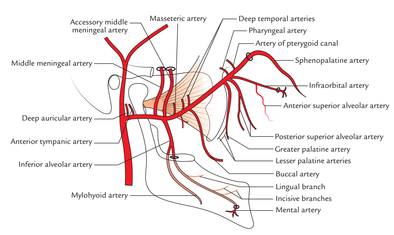
Maxillary Artery: Branches
Branches from the First Part (5 Branches)
1. Deep auricular artery- enters upwards and backwards to goes into the external acoustic meatus by piercing its floor and supplies:
- Skin of external acoustic meatus and
- Outer surfaces of tympanic membrane.
2. Anterior tympanic artery- enters the tympanic cavity by going through petrotympanic fissure and it supplies the inner surface of the tympanic membrane.
3. Middle meningeal artery– is the largest meningeal branch. It supplies meninges along with the skull bone.
Medically it’s the most significant branch of the maxillary artery.
The middle meningeal artery originates from the initial part of the maxillary artery. It ascends upwards deep to the lateral pterygoid, behind the mandibular nerve. Passing between the 2 roots of the auriculotemporal nerve, to goes into the cranial cavity via foramen spinosum in business with meningeal branch of mandibular nerve (nervus spinosus).
As it comes in the cranial cavity, it classes laterally on the floor of the middle cranial fossa and turns upwards and forwards on the greater wing of the sphenoid, where it breaks up into frontal and parietal branches:
- Frontal (anterior) branch, classes upwards in the direction of the pterion and after that arch backwards to ascend in the direction of the vertex, being located over the precentral gyrus of the cerebral hemisphere. In the region of pterion the artery often is located in a bony tunnel in the parietal bone for a centimeter or more.
- Parietal (posterior) branches arch backwards on the squamous part of the temporal bone, cross the lower border of the parietal bone before its mastoid angle; here it splits into branches, which spread out as far as lambda. It is located along the superior temporal gyrus.
Distribution: The middle meningeal artery and its branches are located outside the dura and deep to the inner surface of the skull. These two are supplied by the artery.
The middle meningeal artery and its branches are escorted by corresponding veins, which is located between the artery and the bone.
4. Accessory middle meningeal artery- runs upwards and enters the cranial cavity via foramen ovale.
It supplies meninges and structures in the infratemporal fossa.
5. Inferior alveolar/dental artery- runs downwards between the sphenomandibular ligament and the ramus of the mandible, enters the mandibular foramen, runs via the mandibular canal, supplies molar and premolar teeth and adjoining gingiva. It then breaks up into mental and incisive branches.
The incisive branch supplies the canine and incisor teeth. The mental artery appears via the mental foramen to supply the skin of the chin. Before going into the mandibular foramen the inferior alveolar artery produces 2 branches, specifically.
- Lingual branch: accompanies the lingual nerve and supply the mucous membrane of the cheek.
- Mylohyoid branch: pierces the lower end of the sphenomandibular ligament, enters downwards and forwards to run in the mylohyoid groove. It supplies the mylohyoid muscle.
Branches from the Second Part (4 Branches)
- Deep temporal arteries (generally 2 in number) – ascend upwards on the lateral aspect of the skull deep to the temporalis muscle, which they supply.
- Pterygoid branches- supply the medial and lateral pterygoid muscles.
- Masseteric artery- enters laterally via the mandibular notch and supplies the masseter muscle from its deep surface.
- Buccal artery- supplies buccinator muscle.
Branches from the Third Part (6 Branches)
Posterior superior alveolar artery originates from maxillary artery just before it enters the pterygomaxillary fissure. It breaks up into 2 or 3 branches, which goes into the foramina on the posterior outermost layer of the body of maxilla, runs into alveolar ducts and supplies the molar and premolar teeth and mucus membrane of maxillary air sinus.
Infraorbital artery also appears from maxillary artery just before it reaches the pterygopalatine fossa. The artery enters successively via inferior orbital fissure, infraorbital groove and infraorbital canal and appears on the face via the infraorbital foramen. It supplies these branches:
In the orbit:
- Branches to orbital contents.
- Middle superior alveolar artery to premolar teeth.
- Anterior superior alveolar artery, which descends via canaliculus sinuosus in the anterior wall of the maxillary sinus. It supplies the maxillary air sinus and canine and incisor teeth of the upper jaw.
In the face, it produces branches to supply the lacrimal sac, medial angle of the eye, side of nose and upper lip.
Greater palatine artery enters downwards in the greater palatine canal and appears in the oral cavity in the posterolateral corner of the hard palate via the greater palatine foramen. Now it runs forwards in the groove along the alveolar arch to the incisive fossa where it enters the lateral incisive canal to goes into the nasal cavity. It supplies the roof of the mouth and adjoining gingiva, while in the greater palatine canal the artery produces lesser palatine arteries that appear via foramina of precisely the same name and supply the soft palate and tonsil.
Pharyngeal artery enters backwards via the palatovaginal canal and supplies the mucus membrane of the nasopharynx, auditory tube and the sphenoidal air sinus.
Artery of pterygoid canal runs backwards in the pterygoid canal and supplies the pharynx, auditory tube and the tympanic cavity.
Sphenopalatine artery is regarded as the continuance of the maxillary artery. It’s the most essential branch of the third part of the maxillary artery. It enters the nasal cavity in the posterior part of the superior meatus via sphenopalatine foramen. Here it splits into:
- posterior lateral nasal and
- posterior septal branches.
The posterior lateral nasal branches supply the lateral wall of the nose and sphenoidal and ethmoidal air sinuses, the posterior septal branches cross the undersurface of the body of the sphenoid and after that pass forwards and downwards along the nasal septum. 1 of the branches of this group is long, runs in a groove on the vomer in the direction of the incisive canal and anastomoses with the terminal branch of the greater palatine artery.
Outline
Branches of the maxillary artery.
| First part | Second part | Third part | |
|---|---|---|---|
| Branches | Five | Four | Six |
| 1 | Deep auricular artery | Deep temporal | Posterior superior alveolar (dental) artery |
| 2 | Anterior tympanic artery | Pterygoid branches | Infraorbital artery |
| 3 | Middle meningeal artery | Masseteric artery | Greater palatine artery |
| 4 | Accessory meningeal artery | Buccal artery | Pharyngeal artery |
| 5 | Inferior alveolar artery | Artery of pterygoid canal | |
| 6 | Sphenopalatine artery | ||

 (60 votes, average: 4.87 out of 5)