Temple Fossa, Temporomandibular Joint and Pterygo-palatine Fossa.
The infratemporal fossa is the space underneath the base of the skull, between ramus of the mandible and the side wall of the pharynx. Together via a gap deep to the zygomatic arch it interacts with the temporal fossa. It’s also called the parapharyngeal space or lateral pharyngeal space.
Borders
The bounds of infratemporal fossa are:
- Roof: Created by the temple surface of the higher wing of the sphenoid. It’s pierced by foramen spinosum and foramen ovale. It’s divided from the anterior wall by pterygomaxillary fissure. Created by lateral surface of the lateral pterygoid plate of the sphenoid.
- Medial wall: Created by the ramus of the mandible. Created by the temple outermost layer of the maxilla. It’s divided from roof by inferior orbital fissure.
- Anterior wall: Created by the temple outermost layer of the maxilla. It’s divided from roof by inferior orbital fissure.
- Lateral wall: Created by the ramus of the mandible.
- Posterior wall: Created by styloid process of the temporal bone.
- Floor: Open and goes up to the level of the base of the mandible.
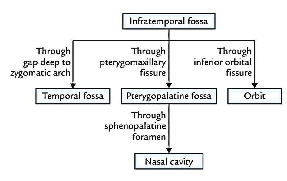
Communications
The infratemporal fossa interacts:
- above with the temporal fossa via a gap, deep to zygomatic arch and middle cranial fossa via foramen ovale and foramen spinosum,
- below it’s constant with the tissue spaces of the neck lateral to the pharynx,
- with the pterygopalatine fossa via pterygo maxillary fissure and
- with the orbit via inferior orbital fissure.
All communications are presented below:
Contents
The major structures existing in the infratemporal fossa are:
- Muscles: Lateral pterygoid, medial pterygoid and tendon of temporalis.
- Blood vessels: Maxillary artery, maxillary vein and pterygoid venous plexus.
- Neural structures: Mandibular nerve, chorda tympani nerve and otic ganglion.
Muscles
Lateral Pterygoid
It’s a short, thick conical muscle with its apex pointing backwards. It enters backwards and somewhat laterally from the roofing and medial wall of the fossa to the neck of the mandible.
Origin
The lateral pterygoid is composed of 2 heads, upper and lower:
- The upper smaller head appears from the temple surface and crest of the higher wing of the sphenoid bone.
- The lower bigger head originates from the lateral surface of the lateral pterygoid plate of the sphenoid bone.
Insertion
The fibres of 2 heads run backwards and laterally and converge to create a thick tendon that is added into:
- Pterygoid fovea on the very front of the neck of the mandible.
- Articular disc and capsule of the temporomandibular joint.
Nerve Supply
Lateral pterygoid is supplied by a branch of anterior section of the mandibular nerve.
Activities
- Lateral pterygoids of 2 sides depress the mandible (opens the mouth) by pulling forwards the condylar processes of the mandible and the articular discs of the temporomandibular joints.
- Medial and lateral pterygoid muscles of 2 sides acting collectively protrude the mandible.
- Medial and lateral pterygoid muscles of both sides contract alternately to create side to side movements of the lower jaw as in mastication.
Key Points
- The lower head of lateral pterygoid enters between both heads of the medial pterygoid muscle.
- It’s the only masticatory muscle, which opens the mouth.
- The articular disc of temporomandibular joint is developmentally a part of tendon of lateral pterygoid muscle.
Connections
The lateral pterygoid is regarded as the key muscle of theinfratemporal region because its relationships supply a reasonable ideaabout the layout of structures in this region. Its connections are:
Superficial:
- Ramus of the mandible.
- Masseter.
- Tendon of temporalis.
- Superficial head of medial pterygoid.
- Maxillary artery and its temporal and masseteric branches.
Deep:
- Mandibular nerve.
- Middle meningeal artery.
- Sphenomandibular ligament.
- Deep head of medial pterygoid muscle.
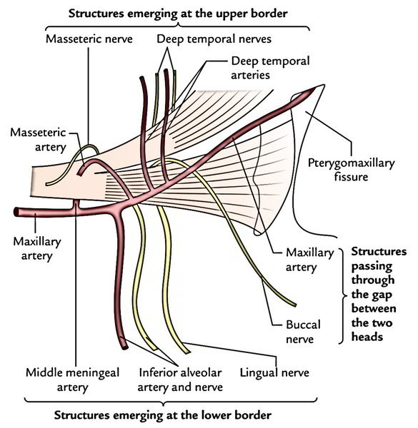
Structures appearing at the upper border:
- Deep temporal nerves (2 in number).
- Masseteric nerve.
Structures appearing at the lower border:
- Inferior alveolar nerve and artery.
- Lingual nerve.
- Middle meningeal artery (it enters up deep to the lower border).
Structures going through the gap between both heads:
- Maxillary artery, which enters the gap to get to the pterygopalatine fossa via pterygomaxillary fissure.
- Buccal nerve, a branch of mandibular nerve. It comes out via the gap to supply sensory innervation to the skin and mucus membrane of the cheek.
Medial Pterygoid
The medial pterygoid is a thick quadrilateral muscle andconsists of 2 heads: superficial and deep.
Origin
The small superficial head (a small slide of muscle) originates from maxillary tuberosity and lateral surface of the pyramidal process of palatine bone.
The large deep head (creating the majority of muscle) originates from medial surface of the lateral pterygoid plate and grooved surface of the pyramidal process of palatine bone.
Insertion
The fibres run downwards, backwards and laterally to be added by a powerful tendinous lamina into a roughened area on the posteroinferior part of the medial surface and angle of ramus of mandible as high as the mandibular foramen and as forwards as the mylohyoid groove.
Nerve Supply
The medial pterygoid is supplied by a nerve to medial pterygoid, a branch from the principal trunk of the mandibular nerve.
Connections
Superficial:
- Lingual nerve.
- Inferior alveolar nerve.
- Inferior alveolar vessels.
Deep:
- Levator palati and tensor palati muscles.
- Superior constrictor of pharynx.
- Styloglossus and stylopharyngeus muscles.
Activities
- Medial pterygoids of 2 sides elevate the mandible to assist in closure of mouth.
- Acting with lateral pterygoids, the medial pterygoids protrude the mandible.
- When medial and lateral pterygoids of 1 side act collectively, the corresponding side of the mandible is rotated forwards and to the opposite side.
- Medial and lateral pterygoids of 2 sides when contract alternately generate side to side movements that are utilized to grind the food.
Blood Vessels
Maxillary Artery

The maxillary artery is the bigger terminal branch of the external carotid artery.
It appears behind the neck of the mandible runs horizontally forwards up to the lower border of lower head of lateral pterygoid. Now it turns upwards and forwards, crosses the lower head of lateral pterygoid superficially (occasionally deep). After coming between the 2 heads of lateral pterygoid it enters the pterygopalatine fossa by going through pterygomaxillary fissure. Here it finishes by supplying its terminal branches.
The maxillary artery has a wide land of distribution. It supplies:
- Upper and lower jaws
- Muscles of temporal and infratemporal fossae
- Nose and paranasal sinuses
- Palate and roof of pharynx
- External and middle ear
- Pharyngotympanic tube and
- Dura mater
The maxillary artery enters the temple fossa by passing forwards, between the neck of mandible and the sphenomandibular ligament.
Maxillary Vein and Pterygoid Venous Plexus
Maxillary Vein
It’s a short venous trunk, which accompanies the initial part of the maxillary artery. It’s created by the confluence of veins from the pterygoid venous plexus and enters backwards between the sphenomandibular ligament and the neck of the mandible. Inside the parotid gland it unifies with all the superficial temporal vein to create the retromandibular vein.
Pterygoid Venous Plexus
It’s a network of quite small veins that are located around and inside the lateral pterygoid muscle.
The pterygoid venous plexus interacts:
- with inferior ophthalmic vein via inferior orbital fissure
- with cavernous sinus by emissary veins via foramen ovale or foramen of Vesalius and
- with facial vein via the deep facial vein
The plexus is emptied by maxillary vein that is created at the lower border of the lateral pterygoid muscle.
The pterygoid venous plexus is occasionally known as a peripheral heart for during yawning when the mouth is broadly open because of contraction of lateral pterygoid muscle. The stagnant venous blood is pumped up into the cavernous sinus and maxillary vein. Perhaps this is actually the reason people yawn in the morning when they get up from slumber.
Neural Structures
The infratemporal fossa includes the mandibular nerve and its branches, chorda tympani nerve and otic ganglion.
Test your Knowledge
Infratemporal Fossa

 (47 votes, average: 4.87 out of 5)