Lumbar Plexus is a network of intersecting nerves in the lumbar region of the human body. The unification of ventral rami of L1 to L3 lumbar nerves and big upper part of ventral ramus of L4 nerve inside the substance of psoas major makes lumbar plexus.
The lumbosacral trunk is created when the lower part of the ventral ramus of L4 nerve joins with all ventral ramus of L5 nerve that is a part of the making of sacral plexus.
Nervi furcalis is the term sometimes used to designate the ramus of the 4th lumbar nerve, because it forms the link between the sacral and lumbar plexuses.
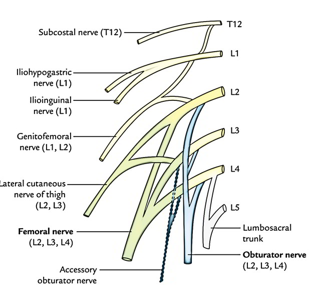
- The ventral ramus of the L1 nerve supplemented by a twig from T12 (subcostal) nerve splits into a bigger upper branch and smaller lower branch. The bigger upper branch supplies rise to iliohypogastric and ilioinguinal nerves.
- The smaller lower branch joins with a twig from the L2 nerve and creates the genitofemoral nerve.
- The L2, L3, L4 nerves break up into dorsal and ventral sections. The dorsal sections of L2, L3, L4 unify to create the femoral nerve. The ventral sections of L2, L3, and L4 join to create the obturator nerve. The accessory obturator nerve if present is originated from the ventral branches of the L3, L4 nerves.
- The main branches of the lumbar plexus are the femoral nerve and the obturator nerve.
Branches of The Lumbar Plexus
| Branches | Root value |
|---|---|
| 1. Iliohypogastric nerve | L1 |
| 2. Ilioinguinal nerve | L1 |
| 3. Genitofemoral nerve | L1, L2 (ventral divisions) |
| 4. Lateral cutaneous nerve of the thigh | L2, L3 (dorsal divisions) |
| 5. Femoral nerve | L2, L3, L4 (dorsal divisions) |
| 6. Obturator nerve | L2, L3, L4 (ventral divisions) |
| 7. Accessory obturator nerve (occasional) | L3, L4 (ventral divisions) |
Course and Distribution of The Branches
Iliohypogastric Nerve
It appears underneath the lateral border of the psoas major muscle, enters downward and laterally in front of the quadratus lumborum. At the lateral border of quadratus lumborum it pierces aponeurotic origin of the transversus abdominis just above the iliac crest and runs in the anterior abdominal wall. It gives cutaneous innervation to the skin of gluteal region and anterior abdominal wall in the hypogastric region.
Ilioinguinal Nerve
It pursues the exact same course as the iliohypogastric nerve, but at a somewhat lower level. It pierces the transversus abdominis near the anterior part of the iliac crest. It gives motor innervation to internal oblique and transversus abdominis muscles and sensory innervation to the skin on the upper medial aspect of the thigh, the root of the penis, and scrotum in the male, and mons pubis and labium majus in the female.
Genitofemoral Nerve
It enters forwards via the psoas, pierces it and its covering psoas fascia. It runs on the anterior outermost layer of the psoas near its medial border and breaks up above the inguinal ligament into genital and femoral branches. The femoral branch runs along the lateral side of the external iliac artery and enters the femoral sheath, where it is located anterolateral to the femoral artery. It pierces the anterior wall of the sheath to supply the skin over the femoral triangle. The genital branch enters the deep inguinal ring and traverses via the inguinal canal along the spermatic cord in the male and the round ligament of uterus in the female. In the male it supplies the cremaster muscle and scrotal skin, and in the female, it supplies the skin of mons pubis and labium majus.
Lateral Cutaneous Nerve of The Thigh (Lateral Femoral Cutaneous Nerve)
It appears underneath the lateral border of the psoas major above the iliac crest, runs downward and laterally across the iliac fossa in front of the iliacus muscle under cover of the iliac fascia. It enters the thigh by passing below the lateral end of the inguinal ligament. Occasionally it goes through the inguinal ligament. It gives cutaneous innervation to the upper lateral aspect of the thigh.
Femoral Nerve
It appears underneath the lateral border of the psoas major below the iliac crest, runs downward and small laterally in the groove between the psoas major and iliacus under cover of the iliac fascia. It enters the thigh by passing deep to the inguinal ligament where it is located lateral to the psoas sheath. Before going into the thigh it supplies the iliacus muscle.
Obturator Nerve
It appears below the medial border of the psoas major in the lumbosacral triangle, crosses the anterolateral angle of the ala of the sacrum to run downward and forward along the lateral wall of the true pelvis, and eventually enters the thigh by going through the obturator canal.
Accessory Obturator Nerve
It’s an inconstant nerve. When present, it accompanies the medial border of the psoas major to go into the thigh. It supplies the pectineus muscles.

 (52 votes, average: 4.69 out of 5)