Penis is the male organ of copulation that is traversed by the urethra gives passage for both urine as well as semen. It overindulges with the blood that makes it a capable of becoming tough and erect. Penis is necessary for entering into the vagina.
Parts
The penis is composed of the following 2 parts:
- Root or radix, a connected portion.
- Body or corpus, a free pendulous portion.
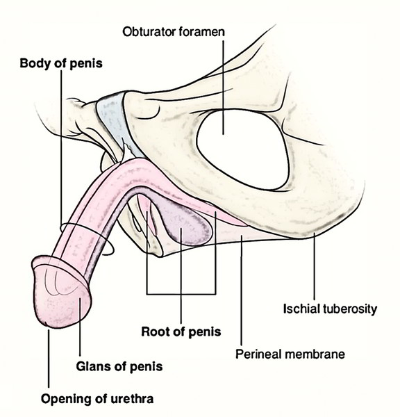
Root (Radix)
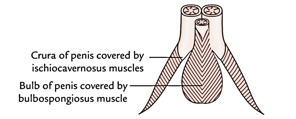
The root of the penis is situated in the superficial perineal pouch and is connected to the inferior aspect of the urogenital diaphragm. It contains 3 masses of erectile tissue, viz. 2 crura (left and right) and the bulb of the penis. Every crus is securely connected to the everted margin of the pubic arch on the corresponding side. Anteriorly, it converges toward its fellow of the opposite side, and near the inferior border of the pubic symphysis the 2 crura come together and continue as the corpora cavernosa of the body of the penis. The bulb is securely connected on the inferior aspect of the perineal membrane extending between the 2 crura. The bulb narrows anteriorly and continues as the corpus spongiosum of the body. Its flattened deep surface above its heart is pierced by the urethra, which traverses its substance to get to the corpus spongiosum. This part of the urethra in the bulb reveals a dilatation in its floor named intrabulbar fossa. Every crus is covered by the ischiocavernosus muscle, and the bulb is covered by the bulbospongiosus muscle.
Body (Corpus)
The body of the penis is the free pendulous portion and is located in front of scrotum. It becomes constant with the root of the penis. In general use, the term penis stands for the body of penis. In the flaccid state, it hangs free below the pubic symphysis, anterior to the scrotum, and ends in an acorn-like enlargement termed glans penis. For convenienceof study and appropriate understanding the body of penis is further split into 3 parts: body appropriate, neck, and glans penis.
Body Appropriate
Shape
In the flaccid state, it’s a long (cylindrical) pendulous structure, and pointed downwards and forwards. In erect state, it’s pointed upwards and forwards and assumes a triangular prism-like shape on cross section with rounded angles. The surfaces of penis are described in this state.
Surfaces
There are 2 surfaces- ventral and dorsal.
- The ventral surface faces backward and downward.
- The dorsal surface faces forward and upward.
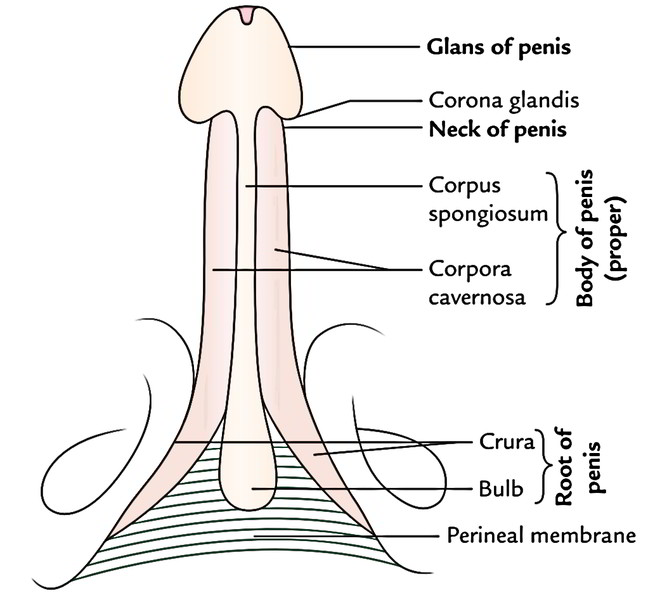
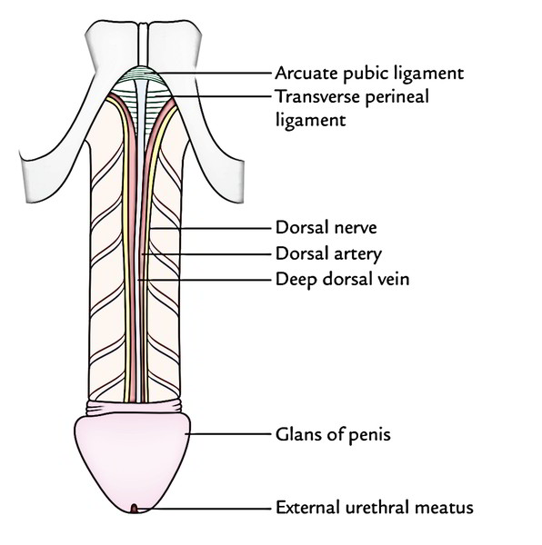
Glans Penis
It’s an enlarged, conical structure in the distal end of the penis, with sagittal slit-like aperture on its peak termed external urethral meatus. The projecting margin of the base of glans is referred to as corona glandis which overhangs the neck of the penis.
Neck of Penis
It’s an obliquely grooved constriction just behind the base of the glans.
Structure
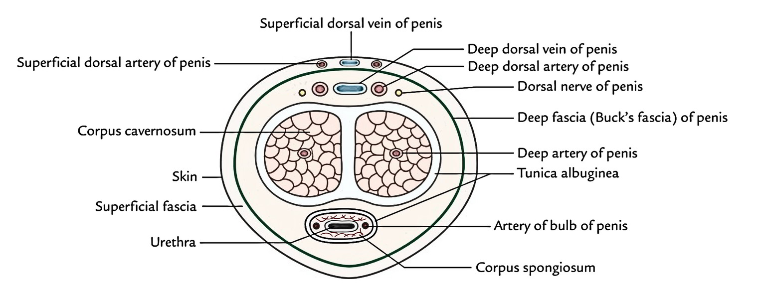
It’s composed of 3 elongated masses of erectile tissue, which are capable of significant enlargement when engorged with blood during an erection. The 3 erectile tissue masses contain 2 corpora cavernosa and 1 corpus spongiosum .
Corpora Cavernosa
Both corpora cavernosa create the greater part of the body of penis. They’re the forward continuance of the crura. Both corpora cavernosa, throughout their length, place in close apposition with 1 another and are encompassed by a common fibrous envelope termed the tunica albuginea. Tunica albuginea is composed of superficial and deep layers. The longitudinally positioned superficial fibres create a tube to enclose both corpora, while the circular deep fibres surround every corpus individually and create at their junction in the midline the septum of penis. This septum is entire proximally but replaced distally by fibrous bands such as the teeth of a comb referred to as pectiniform septum.
The wide groove between the 2 corpora cavernosa on the ventral (urethral) surface lodges the corpus spongiosum. A quite similar but narrower groove on the dorsalsurface lodges the deep dorsal vein of the penis being flanked on every side by the dorsal artery and dorsal nerve of the penis. The corpora cavernosa don’t get to the end of the penis but terminate as conical extremities inside the hollow on the internal aspect of glans penis.
Corpus Spongiosum
- The corpus spongiosum is the forward continuance of the bulb. It’s cylindrical and tapers somewhat toward the distal end where it abruptly grows to create a conical enlargement the glans penis. Throughout its length, it’s traversed by the spongy part of the urethra. It’s also surrounded by a thin fibrous sheath of tunica albuginea.
Coverings
Skin of Penis
It’s unusually thin, fine, dark, and hairless. It envelops the body of penis totally. It’s loosely connected to the fascial sheath of the penis and therefore is freely mobile. At the neck of the penis, it’s folded upon itself to create the prepuce or foreskin, which covers the glans for a varying space. The prepuce is retracted during coitus or manually. The internal layer of prepuce is constant with the thin skin covering the glans securely. The skin covering the glans is constant with the mucous membrane of the urethra in the external urethral orifice. On the urethral aspect of the glans, a small median fold enters from the inner aspect of prepuce to the glans immediately proximal to the external urethral meatus. The susceptibility of the frenulum of prepuce plays an essential function in climax.
Prepuce of The Skin
It’s a fold of skin, which covers the glans for a varying extent and is connected to the neck of the penis.
Frenulum of The Prepuce
It’s a median fold of skin on the ventral surface of glans, which enters from the inner surface of prepuce to the external urethral meatus. It’s highly sensitive.
Preputial Sac
It’s a potential space/cleft between the glans and the prepuce.
Clinical Significance
Medicolegal Importance of Smegma
On the corona glandis and neck of the penis there are numerous small preputial glands (altered sebaceous glands), which secrete sebaceous material named smegma. The smegma has a characteristic smell and accumulates in the preputial sac. It’s of great medicolegal importance in the cases of rape. It ought to be cleaned frequently during washing; otherwise it can cause inflammation and ulceration in the region of the neck of penis.
Superficial Fascia of The Penis
- It includes 2 layers- superficial and deep. The superficial layer is devoid of fat and is composed of loose areolar tissue. It might include few muscle fibres- the peripenic muscle. The deep layer in the lower part of the anterior abdominal wall is condensed and creates the fascia of penis named deep fascia of the penis or Buck’s fascia. It encompasses both corpora cavernosa and corpus spongiosum, but will not go beyond the neck of penis. Buck’s fascia separates the superficial and deep dorsal veins of the penis.
Supports of Penis
The ligaments support the weight of the complimentary pendulous part (body) of the penis. They’re 2 in number.
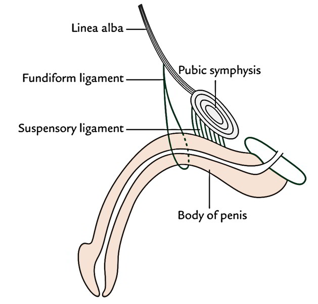
Fundiform Ligament
It springs from the lower part of the linea alba and splits into 2 lamellae, which enclose the proximal part of the body of penis and after that connect on its urethral aspect together with the septum of scrotum.
Suspensory Ligament
It’s deep to the fundiform ligament and triangular in shape. Its narrow upper end is connected in front of the pubic symphysis and broad lower part mixtures with Buck’s fascia (fascia of penis) on either side of the body of penis.
Arterial Supply
The penis is provided by the subsequent 4 pairs of arteries:
- Deep arteries of the penis.
- Dorsal arteries of the penis.
- Arteries of the bulb.
- Superficial dorsal arteries of penis.
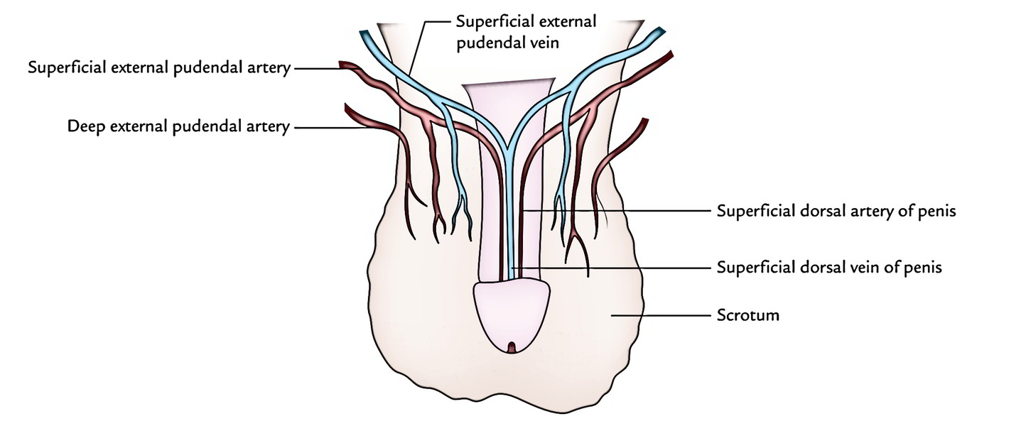
Out of these, the very first 3 pairs of arteries originate from internal pudendal arteries, branches of anterior sections of internal iliac arteries, while the last pair originates from superficial external pudendal arteries, branches of femoral arteries.
The penis is provided mainly by deep arteries and dorsal arteries. Deep arteries are the main vessels for filling the lacunae of erectile tissue during erection of the penis. Every deep artery of the penis runs lengthwise in corpus cavernosum and produces numerous branches. These arteries generate tiny arteries which directly open into the cavernous spaces. In the flaccid state of the penis, these vessels project in the lacunae as looped or spiral vessels and therefore named “helicine arteries.” The dorsal arteries run on the dorsum below the deep fascia and provide the glans penis and distal part of corpus spongiosum, prepuce, and frenulum of prepuce. The arteries of bulb provide the bulb and proximal half of the corpus spongiosum. The superficial external pudendal arteries provide the skin and fascia of the penis.
Venous Drainage
The following 2 veins primarily drain the venous blood from the penis:
- Superficial dorsal vein of the penis.
- Deep dorsal vein of the penis.
The main veins contrary to the arteries be located in the midline divided from every other by the deep (Buck’s) fascia of penis.
Superficial Dorsal Vein of The Penis
It is located in the superficial fascia on the dorsal aspect of the penis in the midline and is generally visible via the skin. Proximally, it splits into left and right branches, which drain into the superficial external pudendal veins.
Deep Dorsal Vein of The Penis
It is located deep to the deep fascia of penis in the median plane dorsally in the narrow groove between both corpora cavernosa.
Deep vein empties majority of the venous blood from the cavernous tissue. Proximally it goes through the gap between the perineal membrane and the lower border of the pubic symphysis to drain into the prostatic venous plexus. The other veins correspond to the arteries.
Lymphatic Drainage
The lymphatic drainage is significant due to its role in metastasis of penis cancer, which takes place often. Lymph vessels from the glans penis drain into the deep inguinal lymph nodes, particularly into the lymph node of Cloquet. The lymph vessels from the remainder of penis drain into superficial inguinal lymph nodes.
Nerve Supply
Sensory innervation: Sensory provide to the penis is originated from the following:
- Dorsal nerve of penis.
- Ilioinguinal nerve.
Motor innervation: The muscles of penis are provided by the perineal branch of pudendal nerve.
Autonomic innervation: The autonomic nerves of the penis are originated from the pelvic (inferior hypogastric) plexus through the prostatic plexus. The sympathetic nerves are vasoconstrictor while parasympathetic fibres areasodilator. The parasympathetic fibres (nervi erigentes) are originated from S2, S3, and S4 spinal sections. The autonomic fibres are spread via the pudendal nerve.
Mechanism of Erection And Ejaculation
The mechanism of erection of penis is only a vascular phenomenon and takes place in response to the parasympathetic excitement. The sexual arousal results in accelerated inflow of blood from helicine arteries in the cavernous spaces of erectile tissue of the penis. The filling of blood in the cavernous spaces leads to the compaction of the veins, which drain erectile tissues. (This is connected with simultaneous spasm of venous smooth muscle sphincters.) Since the venous outflow is occluded, the blood pressure in the cavernous spaces causes inflation and rigidity of the erectile tissue referred to as erection. The pressure inside the corpora cavernosa is 100 mmHg during the time of penile erection.
Points to be noticed
- The strong fibrous envelope (tunica albuginea) significantly contributes to the rigidity of the engorged corpora.
- Erection is controlled by nervi erigentes (S2 S4).
- The ejaculation is composed of 2 processes- discharge and ejaculation. The discharge is the transmission of seminal fluid from vasa deferentia, seminal vesicles, and prostate into the prostatic urethra. The ejaculation is the onward transmission of seminal fluid from the prostatic urethra to the outside.
The sympathetic outflow from T11 to L2 spinal sections results in discharge of semen by causing constriction of smooth muscle of vasa deferentia, seminal vesicles, and prostate. It also constricts the internal urethral opening to stop the retrograde flow of semen into the bladder.
- The rhythmic contraction of bulbospongiosus, provided by the perineal nerve (somatic), compresses the penile urethra and expels the fluid to the outside- ejaculation.
The nervous commands of erection and ejaculation are distinct; erection is mediated by parasympathetic nerve fibres on the other hand ejaculation is mediated by sympathetic and somatic nerve fibres.
Clinical Significance
Impotence
The failure to attain tumescence erection is termed impotence. The commonest causes of erectile dysfunction are: (a) psychogenic interference with failure to relax the smooth muscle in the corpora, (b) arterial insufficiency due to atheromatous disease, and (c) participation of nervi erigentes (parasympathetic outflow) secondary to diabetes.
Priapism
The persistant erection is referred to as priapism. It happens because of persistant spasm of venous smooth muscle sphincters.
Peyronie’s Disease/Chordee
It appears because of a localized thickening or plaque of the corpora cavernosa, which prevents growth of a section of erectile tissue during erection.
Consequently penis becomes arch particularly during erection.
Phimosis
It’s a narrowing of the distal end of the prepuce (foreskin), which prevents its retraction over the glans penis and might interfere together with the micturition (passage of urine).
Paraphimosis
It’s an unusual condition where narrowing of the prepuce is inadequate to interfere together with the micturition, but the prepuce is just enough tight to get stuck on the glans posteriorly on erection and consequently interfere with copulation.
Circumcision
It’s the surgical removal of the prepuce (foreskin of the penis). In kids and grownups, the circumcision is occasionally required to alleviate the patient from a closely constricting prepuce (phimosis). The rite of circumcision for religious motives is one of the earliest surgical processes in the world.

 (61 votes, average: 4.34 out of 5)