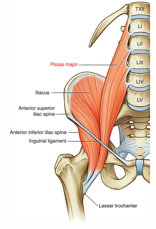
Psoas Major is a long fusiform muscle stretching from the sides of lumbar vertebrae to the lesser trochanter of the femur. The enclosing of the Psoas major is a fascial sheath known as Psoas Sheath.

Psoas Major
Origin
The origination of the psoas major muscle takes place from 14 fleshy skids, which are as follows:
- 5 chemises from intervertebral discs between T12 L5 vertebrae and adjoining margins of the bodies of these vertebrae.
- 5 slides from anterior surfaces and lower edges of the transverse process of 5 lumbar vertebrae.
- 4 skids from tendinous arches bridging the constricted sides of the bodies of lumbar vertebrae. The lumbar vessels pass deep to these arches.
Insertion
From its origin site, the muscle which descends along the pelvic brim, enters the thigh behind the inguinal ligament. Below the ligament, the tendon creates on the lateral side of the muscle, enters in front of the hip joint, and enters the anterior surface of the tip of the lesser trochanter of the femur. A synovial bursa that might communicate together with the cavity of the hip joint normally separates this tendon from the joint capsule.
Nerve Supply
The psoas major is innervated by the branches from ventral rami of L2, L3, L4 spinal nerves.
Actions
Standard Mover Actions
- Flexes the thigh at the hip joint.
- Posteriorly tilts the pelvis at the LS joint.
- Laterally rotates the thigh at the hip joint.
- Elevates the same-side pelvis at the LS joint.
Reverse Mover Actions
- Flexes the trunk at the spinal joints.
- Anteriorly tilts the pelvis at the hip joint.
- Contralaterally rotates the trunk and pelvis.
- Laterally flexes the trunk at the spinal joints.
Functions
Eccentric Antagonist Functions
- Restrains thigh extension and pelvic posterior tilt at the hip joint.
- Restrains thigh medial rotation and pelvic ipsilateral rotation at the hip joint.
- Restrains lumbar spinal extension.
- Restrains lumbar spinal lateral flexion to the opposite side.
- Restrains ipsilateral rotation of the lumbar spine.
- Restrains anterior tilt and depression of the same-side pelvis at the LS joint.
Note
A force of lateral rotation of the thigh at the hip joint prevents medial rotation of the entire lower extremity (thigh, leg, and talus) if the weight-bearing foot overly pronates (if the arch of the foot drops).
Isometric Stabilization Functions
- Stabilizes the lumbar spine at the spinal joints.
- Stabilizes the pelvis at the LS and hip joints.
- Stabilizes the thigh at the hip joint.
Note
Because the psoas major lies so close to the lumbar spine, its major stabilization function is at the spine.
Relations

Psoas Major: Relations
The psoas major is the key muscle of the posterior abdominal wall, because its relationships supply a rational notion about the layout of structures in this region:
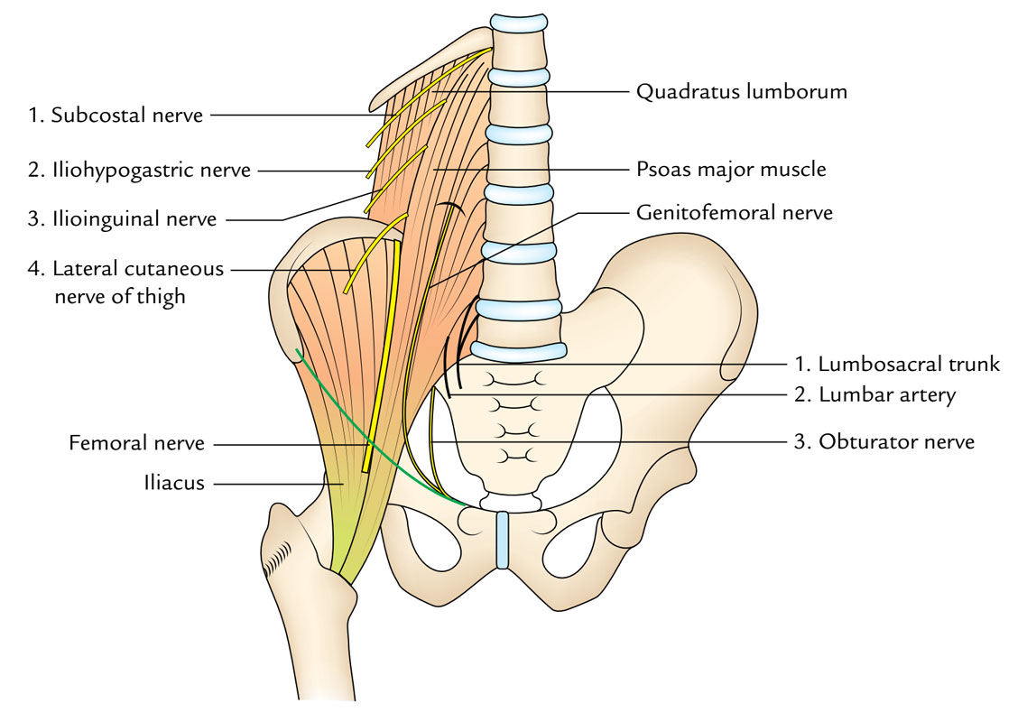
Lumbar plexus creates inside the substance of psoas. The plexus can be shown only by ripping the muscle, for it will not break up the muscle into planes.
5 nerves appear from underneath the lateral border of the psoas major from above downward; these are as follows:
- Subcostal nerve.
- Iliohypogastric nerve.
- Ilioinguinal nerve.
- Lateral cutaneous nerve of thigh.
- Femoral nerve.
The upper 4 nerves come above the iliac crest and runs downward and laterally across the quadratuslumborum muscle. The final nerve (femoral nerve) comes below the iliac crest and runs down in the groove between the psoas and iliacus muscles.
1 nerve (genitofemoral nerve) runs downward on the very front of the psoas major and occasionally may be mistaken for the tendon of psoas minor muscle.
3 significant structures being located on the medial side of the psoas major. From medial to lateral side these are: (a) lumbosacral trunk, (b) iliolumbar artery, and (c) obturator nerve.
Lumbosacral Triangle of Marcille

Psoas Major: Lumbosacral Triangle of Marcille
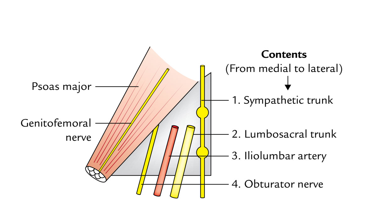
Psoas major’s a triangular separation on every side of the body of L5 vertebra with all the apex directed upward. It’s bounded medially by the body of L5 vertebra, laterally by the medial border of the psoas major, and inferiorly (base) by the ala of the sacrum. The apex is composed by the junction of psoas and the body of L5 vertebra. The floor (posterior wall) is composed by the transverse process of L5 vertebra and iliolumbar ligament.
It includes 4 structures. From medial to lateral sides, these are:
- Sympathetic trunk.
- Lumbosacral trunk.
- Iliolumbar artery.
- Obturator nerve.
Psoas Sheath
The psoas major muscle is enclosed in a fascial sheath named (psoas sheath) created by the psoas fascia. The attachmentsof psoas fascia are as follows:
- Above: It’s thickened to create medial arcuate ligament, which goes from the body of L1 vertebra to the tip of its transverse process.
- Laterally: It combines with all the anterior layer of the thoracolumbar fascia.
- Medially: It’s connected to the bodies and intervening intervertebral discs of lumbar vertebrae and presents 4 tendinous arches.
- Below: It fuses with the arcuate line of the pelvis and the fascia covering the iliacus muscle (iliac fascia).
Clinical Significance
Psoas Abscess

Psoas Major: Psoas Abscess
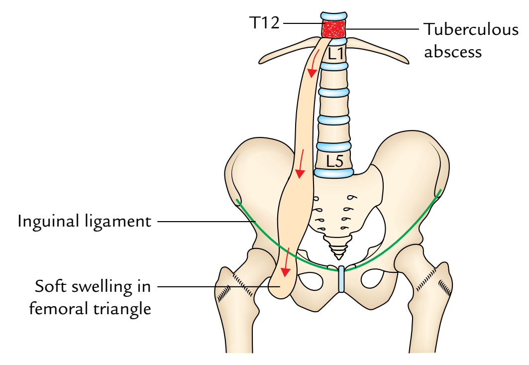
The psoas fascia creates a long tubular sheath (osseofibrous tunnel) termed psoas sheath going from the upper lumbar region of the posterior abdominal wall to the groin. Tubercular infection of vertebrae of the thoraco-lumbar region causes destruction of their bodies resulting in the formation of an abscess. The pus can not propagate anteriorly as a result of anterior longitudinal ligament. For that reason, it propagates laterally into the psoas sheath creating psoas abscess. The pus can also goes into the psoas sheath from the posterior mediastinum via a gap deep to medial arcuate ligament. Pus may subsequently propagate downward along the psoas muscle, below the inguinal ligament into the femoral triangle where it creates a soft swelling.
Palpation
- With the client supine with a pillow under the knees, place palpating hand on the abdomen between the iliac crest and the twelfth rib (lateral to the rectus abdominis, approximately halfway between the ASIS and the umbilicus).
- Palpate slowly but deeply with even pressure directed posteromedially toward the spine.
- Ask the client to flex the thigh at the hip joint a few degrees and feel for the contraction of the psoas major.
- The distal belly of the psoas major is also palpable in the proximal anterior thigh between the pectineus and the sartorius. Note: Be careful with palpation in this region because the femoral nerve, artery, and vein lie over the iliopsoas and pectineus here.

 (58 votes, average: 4.81 out of 5)