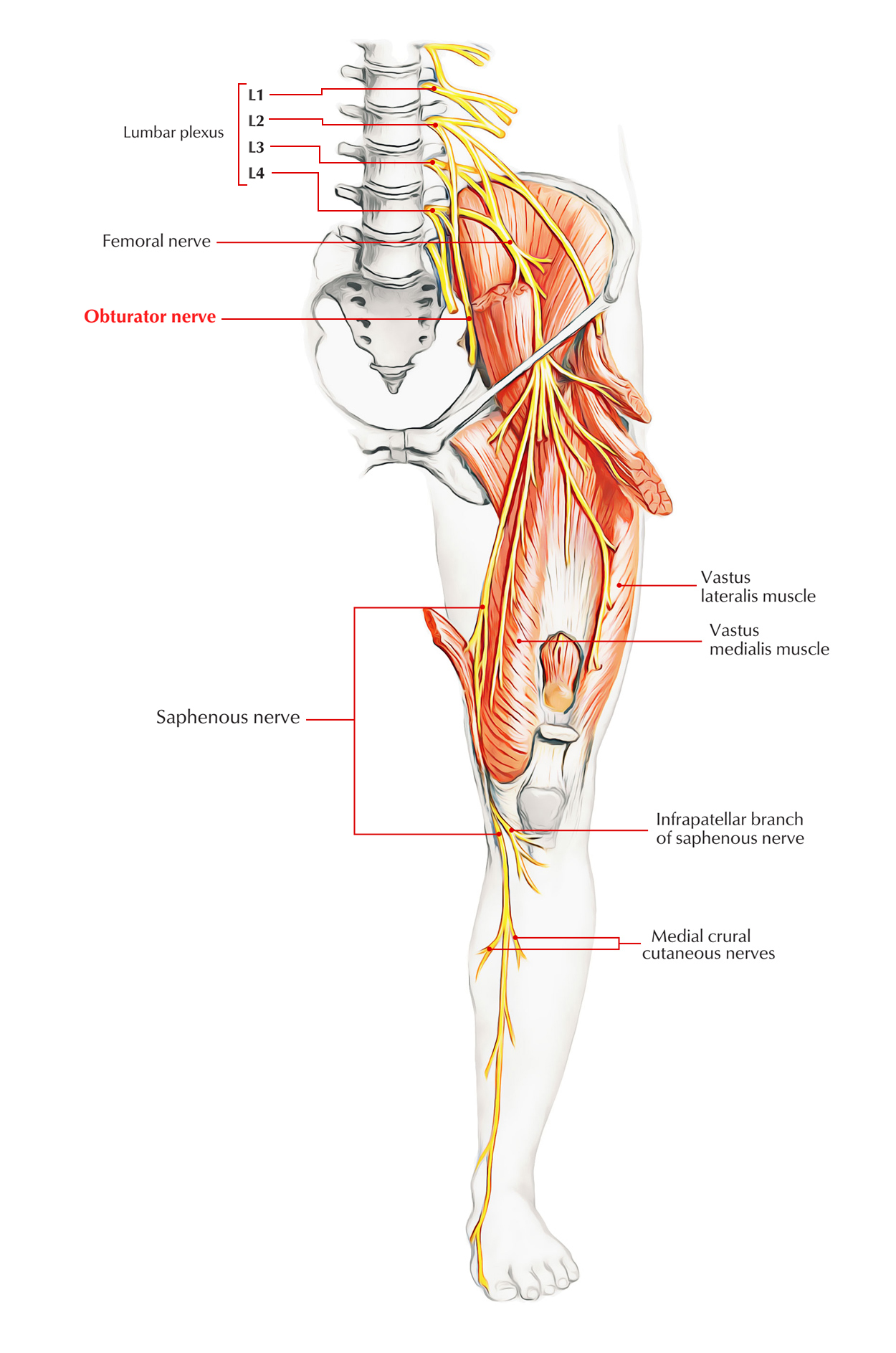
Obturator Nerve belongs to the adductor compartment of the thigh. The inside of psoas major from anterior sections of the ventral rami of L2 to L4 spinal nerves. The nerve goes down in psoas major and issues from its medial border at the ala of the sacrum. It runs through the upper anterior part of the obturator foramen to the medial (adductor) compartment of the thigh while descending along the lateral wall of the lesser pelvis on the obturator internus. It divides into anterior and posterior which stride the adductor brevis muscle, close to the obturator foramen. All muscles of the adductor compartment of thigh are supplied by its motor branches. Cutaneous area on the lower-half of the medial aspect of thigh are supplied by its sensory branches. Articular branches to the hip and knee joints are also supplied by it.

Obturator Nerve
Course and Distribution
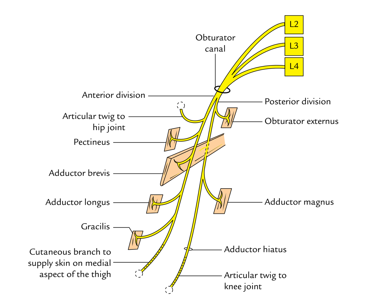
While going through the obturator canal the obturator nerve divides into anterior and posterior sections.

Obturator Nerve: Course and Distribution
1. The anterior section enters downwards into the thigh in front of the obturator externus. It then descends behind the pectineus and the adductor longus, and in front of the adductor brevis. The anterior section supplies the following muscles:
- Pectineus.
- Adductor longus.
- Gracilis.
- Adductor brevis.
The anterior section also supplies an articular twig to the hip joint. Distal to the adductor longus, it enters the adductor canal where it gives a twig to the subsartorial plexus of nerves and ends by supplying the femoral artery in the adductor canal.
2. The posterior section enters the thigh by piercing the anterior part of the obturator externus muscle which it supplies. It then descends behind the adductor brevis and in front of the adductor magnus. The posterior section supplies the following muscles:
- Obturator externus.
- Adductor magnus.
- Adductor brevis.
Its terminal part creates an articular branch termed genicular branch, which pierces the adductor magnus or goes through hiatus for femoral vessels to reach the popliteal fossa where it runs along the popliteal vessels and pierces the oblique popliteal ligament to supply the knee joint.
Clinical Significance
Injury of the Obturator Nerve
The obturator nerve could possibly be injured in the anterior dislocation of the hip joint, orduring radical retropubic prostatectomy. Listed here are the characteristic clinical features:
- Motor reduction: Decline of adduction of the thigh, because of paralysis of adductor muscles of the thigh.
- Sensory loss: Sensory loss on the medial aspect of the thigh, because of participation of the cutaneous branch of the anterior section of the obturator nerve.
Obturator Nerve Neuropathy
The syndrome of an obturator nerve entrapment causing the medial thigh pain is described in sportsmen with large adductor muscles.
Surgical Section of the Obturator Nerve
It’s sometimes carried out to alleviate the spasm of adductor muscles in the spastic paralysis.
Annoyance of the Obturator Nerve
The inflammation of the ovary causes localised peritonitis in the region of ovarian fossa which might cause annoyance of the obturator nerve. In this case, the pain could possibly be referred to the hip, knee, and medial side of the thigh.
Referred Pain
In diseases of the hip joint, the pain could be referred to the medial side of the thigh.
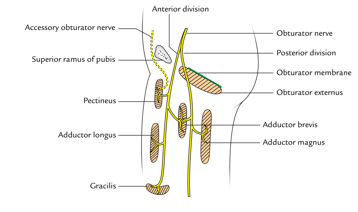
Accessory Obturator Nerve

Obturator Nerve: Accessory Obturator Nerve
In about 30% people, accessory obturator nerve originates from the lumbar plexus. It’s created by the ventral sections of the anterior primary rami of L3, L4 spinal nerves. It crosses the superior ramus of pubis deep to pectineus, which it furnishes. It supplies an articular twig to the hip joint and ends by communicating with all the anterior section of the obturator nerve. If small, it supplies only the pectineus muscle.

 (55 votes, average: 4.67 out of 5)