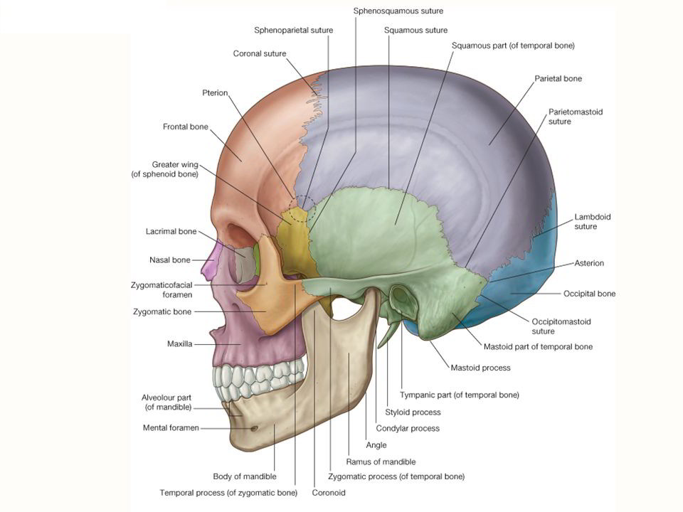
The norma lateralis includes the lateral wall of the skull and the following bones
- Frontal
- Parietal
- Occipital
- Temporal
- Sphenoid
- Zygomatic
- Mandible
- Maxilla
- Nasal
These bones form norma lateralis.
Features
Temporal Lines
The temporal lines have been studied in the norma verticalis. The inferior temporal line, in its posterior part, turns downwards and forwards and becomes continuous with the supramastoid on the squamous temporal bone near its junction with the mastoid temporal. This crest is continuous anteriorly with the posterior root of the zygoma.
Zygomatic Arch
The zygomatic arch is a horizontal bar on the side of the head, in front of the ear, a little above the tragus. It is formed by the temporal process of the zygomatic bone in anterior one-third and the zygomatic process of the temporal bone in posterior two-thirds. The zygomatico-temporal crosses the arch obliquely downwards and backwards.
The arch is separated from the side of the skull by a gap which is deeper in front than behind. Its lateral surface is subcutaneous. The anterior end of the upper border is called the jugal point. The posterior end of the zygoma is attached to the squamous temporal bone by anterior and posterior roots. The articular tubercle of the root of the zygoma lies on its lower border, at the junction of the anterior and posterior roots. The anterior root passes medially in front of the articular fossa. The posterior root passes backwards along the lateral margin of the mandibular fossa, then above the external acoustic meatus to become continuous with the supramastoid crest. Two projections are visible in relation to these roots. One is articular tubercle or tubercle of the root of zygoma at its lower border. The other is visible just behind the mandibular or articular fossa and is known as postglenoid tubercle.
External Acoustic Meatus
The external acoustic meatus opens just below the posterior part of the posterior root of the zygoma. Its anterior and inferior margins and the lower part of the posterior margin are formed by the tympanic plate and the posterosuperior margin is formed by the squamous temporal bone. The margins are roughened for the attachment of the auricular cartilage.
The suprameatal triangle is a small depression posterosuperior to the meatus. It is bounded above by the supramastoid crest, in front by the posterosuperior margin of the external meatus and behind by a vertical tangent to the posterior margin of the meatus. The suprameatal spine may be present on the anteroinferior margin of the triangle. The triangle forms the lateral wall of the tympanic or mastoid antrum.
Mastoid Part
The mastoid part of the bone lies just behind the external acoustic meatus. It is continuous anterosuperiorly with the squamous temporal bone. A partially obliterated squamomastoid suture may be visible just in front of and parallel to the roughened area for muscular insertions.visible just in front of and parallel to the roughened area for muscular insertions.
The mastoid temporal bone articulates posterosuperiorly with the posteroinferior part of the parietal bone at the horizontal parietomastoid suture: and posteriorly with the squamous occipital bone at the occipitomastoid suture. These two sutures meet at the lateral end of the lambdoid suture. The asterion is the point where the parietomastoid, occipitomastoid and lambdoid sutures meet. In infants the asterion is the site of the posterolateral or mastoid fontanelle, which closes at the end of the first year.
The mastoid process a nipple-like large projection from the lower part of the mastoid temporal bone, posteroinferior to the external acoustic meatus. It appears during the second year of life. The tympanomastoid fissure is placed on the anterior aspect of the base of the mastoid process. The mastoid foramen lies at or near the occipitomastoid suture.
Styloid Process
The styloid process is a needle like thin, long projection from the norma basalis situated anteromedial to the mastoid process. It is directed downwards, forwards and slightly medially. Its base is partly ensheathed by the tympanic plate. The apex or tip is usually hidden from view by the posterior border of the ramus of the mandible.
Temporal Fossa
The Boundaries of the temporal fossa are as follows:
- Above, by the temporal line of the frontal bone.
- Below, by the upper border of the zygomatic arch laterally; and by the infratemporal crest of the greater wing of the sphenoid bone medially. Through the gap deep to the zygomatic arch, the temporal fossa communicates with the infratemporal fossa.
- The anterior wall is formed, by the zygomatic bone and by parts of the frontal and sphenoid bones. This wall separates the fossa from the orbit.
The Floor of the temporal fossa are as follows:
- The anterior part of the floor is crossed by an H-shaped suture where four bones; frontal, parietal, sphenoid and temporal adjoin each other
- This area is termed the pterion. It lies 4 cm above the midpoint of the zygomatic arch or 4 cm above the zygoma and 2.5 cm behind the frontozygomatic suture. Deep to the pterion there lie the middle meningeal-vein, the anterior division of the middle meningeal artery, and the stem of the lateral sulcus of the brain.
- On the temporal surface of the zygomatic bone forming the anterior wall of the fossa there is the zygomatico- temporal foramen.
Infratemporal fossa
The walls are divided in four parts medial, lateral, anterior and posterior.
- The medial wall is formed by the lateral pterygoid plate and the pyramidal process of the palatine bone.
- The lateral wall is formed by the ramus of the mandible.
- The anterior wall is formed by the infratemporal or posterior surface of the maxilla and by the medial surface of the zygomatic bone. The anterior and medial walls are separated in their upper parts by the pteiygomaxillary fissure through which the infratemporal fossa communicates with the pterygopalatine fossa. The upper end of the pterygomaxillary fissure is continuous with the anterior part of the inferior orbital fissure through which the infratemporal fossa communicates with the orbit.
- The posterior wall is open.
The roof is formed medially by the infratemporal surface of the greater wing of the sphenoid and by a small part of the squamous temporal bone. Laterally, the roof is incomplete where the infratemporal fossa communicates with the temporal fossa through the gap deep to the zygomatic arch. The roof formed by greater wing is pierced by the foramen ovale and by the foramen spinosum.
The floor of norma lateralis is open.

 (52 votes, average: 4.77 out of 5)