Body cavities are spaces inside the body which contain, protect, separate, and support internal organs. There are two major cavities of the body which contain internal organs: the dorsal (posterior) and ventral (anterior) cavities. The body cavities protect as well as cushion the contained organs and permit changes in their size and shape without affecting surrounding tissues.
Dorsal Cavity
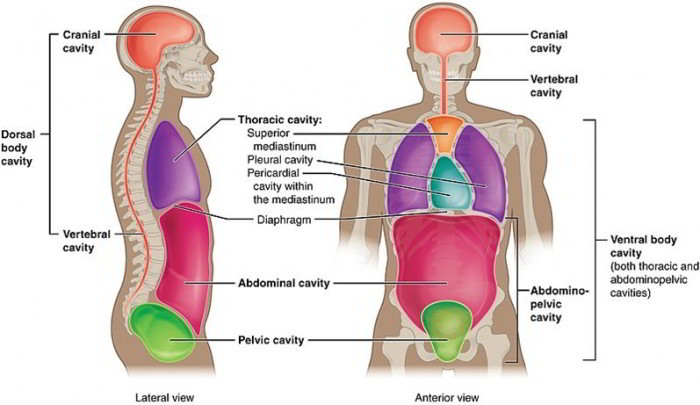
The dorsal cavity is subdivided into the cranial cavity, which houses the brain, and the vertebral canal, which in turn contains the spinal cord. Observe in the figure mentioned above how the cranial bones and the vertebral column form the walls of the dorsal cavity and offer protection for these delicate organs.
Ventral Cavity
The ventral cavity is divided by the diaphragm, a thin dome-shaped sheet of muscle, into a superior thoracic cavity as well as an inferior abdominopelvic cavity. The thoracic cavity is guarded by the rib cage and contains the heart and lungs. The abdominopelvic cavity is subdivided into a superior abdominal cavity and an inferior pelvic cavity, however, there is no structural separation between them. To visualize the separation, think of a transverse plane passing through the body just superior to the pelvis. The abdominal cavity includes the stomach, intestines, liver, gallbladder, pancreas, spleen, and kidneys. The pelvic cavity includes the urinary bladder, sigmoid colon, rectum, and internal reproductive organs.
Membranes of Body Cavities
The membranes lining body cavities help and protect the internal organs in the cavities.
Dorsal Cavity Membranes
The dorsal cavity is lined by 3 layers of protective membranes which are collectively termed the meninges. One of the most superficial membrane is connected to the wall of the dorsal cavity, and the deepest membrane tightly covers the brain and spinal cord.
Ventral Cavity Membranes
The ventral body cavity organs are supported as well as protected by serosae (singular, serosa), or serous membranes. The serous membranes are thin layers of tissue that line the body cavity and protect the internal organs. Serous membranes have a superficial parietal layer which lines the cavity and a deep visceral layer which covers the organ. The parietal and visceral layers secrete a watery lubricating fluid that is generically referred to as serous fluid into the cavity made between the layers. This setup is similar to that of a fist pushed into a balloon (The serous membranes of the body are the pleura, pericardium, and peritoneum.
The serous membranes lining the thoracic cavity are named pleurae (singular, pleura), or pleural membranes. The walls of the left and right portions of the thoracic cavity are lined by the parietal pleurae. The surfaces of the lungs are covered by the visceral pleurae. The parietal and visceral pleurae are separated by a thin film of serous fluid named pleural fluid, which decreases friction as the pleurae rub against one another as the lungs expand and contract during breathing. The potential space (not an actual space) between the parietal and visceral pleurae is referred to as the pleural cavity.
The left and right portions of the thoracic cavity are divided by a membranous partition, the mediastinum. Organs situated within the mediastinum consist of the heart, thymus, esophagus, and trachea.
The heart is wrapped up by the pericardium which is formed by membranes of the mediastinum. The thin visceral pericardium is tightly stuck to the surface of the heart. The parietal pericardium lines the deep surface of a loosely fitting sac around the heart. The potential space between the visceral and parietal pericardia is the pericardial cavity, moreover it contains serous fluid, termed pericardial fluid, that decreases friction as the heart contracts and relaxes. The walls of the abdominal cavity and the surfaces of abdominal organs are lined with the peritoneum, or peritoneal membrane. The parietal peritoneum lines the walls of the abdominal cavity but not the pelvic cavity. It comes down only to cover the superior portion of the urinary bladder.
The kidneys, pancreas, and parts of the intestines lie posterior to the parietal peritoneum in a space referred to as the retroperitoneal space. The visceral peritoneum, an extension of the parietal peritoneum, covers the surface of the abdominal organs. Double layered folds of the visceral peritoneum, the mesenteries, extend between the abdominal organs and offer support for them. The potential space between the parietal and visceral peritoneal membranes is termed as the peritoneal cavity and includes a small amount of serous fluid referred to as peritoneal fluid.

 (52 votes, average: 4.52 out of 5)