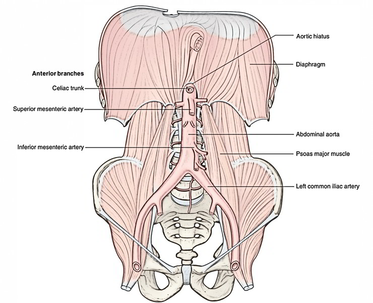
Abdominal Aorta is the main blood vessel in the abdominal cavity. It is the largest blood vessel in the abdomen opposite to the lower border of the T12 vertebra or intervertebral disc between vertebrae T12 and L1 that starts as the continuance of descending thoracic aorta in the aortic orifice of the diaphragm is known as the abdominal aorta. It descends vertically downward and somewhat to the left, in front of the vertebral column, and ends in front of the lower part of the body of L4 vertebra (about 1.25 cm) to the left of the median plane by splitting into left and right common iliac arteries. It keeps on diminishing in size rapidly due to the use of many large branches. Due to its dependence on the bodies of the vertebrae, it has a convex forward curve. The summit of its convexity corresponds to the third lumbar vertebra.
Measurements
- Length: 10-11 cm.
- Width: 2 cm.
Relations
Posterior: Abdominal aorta it is separated into:
- Bodies of the upper 4 lumbar vertebrae and intervening intervertebral discs.
- Anterior longitudinal ligament.
- Third and fourth left lumbar veins.
Anterior: From above downward, the aorta is linked to these structures:
- Branches of the celiac artery and the celiac plexus.
- Aortic plexus.
- Pancreas and splenic vein.
- Left renal vein.
- Third part of the duodenum.
- Root of the mesentery.
- Coils of the small intestine divided by parietal peritoneum.
Right side:
- The upper part of the inferior vena cava is separated from azygos vein, cisterna chyli, thoracic duct, and the right crus of the diaphragm and from the right celiac ganglion;
- The aorta and inferior vena cava are connected .
Left side:
- Left sympathetic trunk.
- Left crus of the diaphragm.
- Left celiac ganglion.
- The ascending part of the duodenum.
Branches
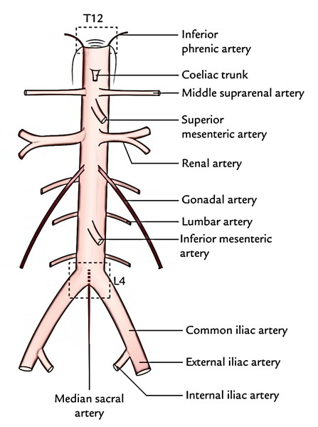
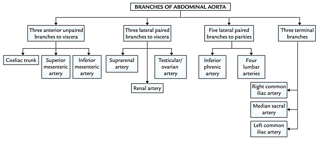
The abdominal aorta supplies 3 sets of branches:
- 3 unpaired ventral branches to the gut.
- 3 coupled lateral branches to 3 matched glands (suprarenal glands, kidneys, and gonads).
- Coupled posterolateral branches to the abdominal wall.
Along with the above, the aorta also supplies rise to coupled inferior phrenic artery, unpaired median sacral artery, and 2 terminal branches.
Coeliac Trunk (Celiac Artery)
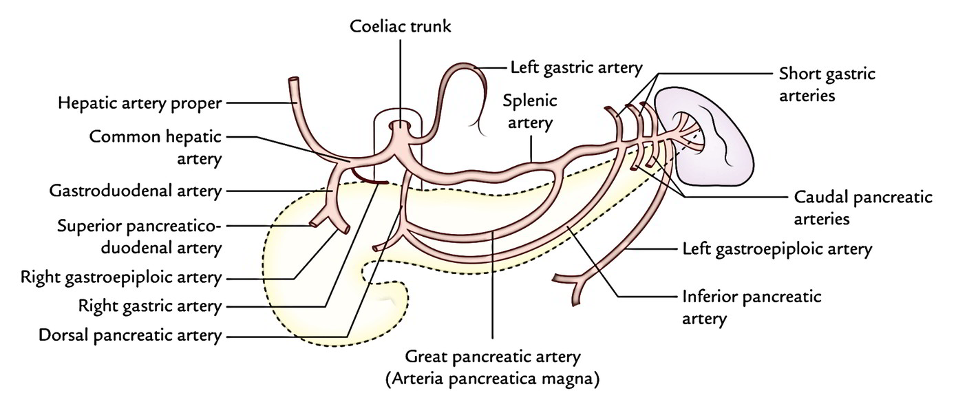
It’s a short, wide vessel (1.25 cm long), which originates from the front of the abdominal aorta immediately below the aortic opening of the diaphragm at the level of the intervertebral disc between T12 and L1 vertebrae. It runs forwards and somewhat to the right and immediately breaks up into following 3 branches: (a) left gastric artery, (b) common hepatic artery, and (c) splenic artery.
Left Gastric Artery (Smallest Branch)
It enters upward and to the left to get to the cardiac end of the stomach where it turns forwards to run downward along the lesser curvature of the stomach. It supplies rise to esophageal branches at the cardiac end of the stomach and numerous gastric branches along the lesser curvature of the stomach.
Common Hepatic Artery
It’s bigger in relation to the left gastric artery. It runs downward, forward, and to the right to make it to the upper surface of the very first part of the duodenum. Here it turns upward and runs in the right free margin of the lesser omentum (as hepatic artery appropriate) in front of the portal vein and to the left of common bile duct. On reaching the porta hepatis, it ends by dividing into left and right hepatic arteries.
The common hepatic artery supplies rise to these branches:
- Gastroduodenal artery appears at the upper border of the first of the duodenum. It runs downward behind the duodenum and ends at its lower border by splitting into right gastroepiploic and superior pancreatic duodenal arteries.
- Right gastric artery originates at just distal to the origin of gastroduodenal artery. It turns to the left and runs upward along the lesser curvature of the stomach.
- Hepatic artery appropriate is the continuance of the common hepatic artery. It supplies rise to left and right hepatic arteries at the porta hepatis. The right hepatic artery supplies origin to cystic artery to supply the gallbladder. The left and right hepatic arteries supply the left and right physical lobes of the liver, respectively.
Splenic Artery (Largest Branch)
It’s unusually tortuous. It runs horizontally to the left, along the upper border of the body and tail of the pancreas, crosses in front of left suprarenal gland and anterior surfaceof the upper part of kidney to goes into the lienorenal ligament via which it reaches the hilum of spleen. Here it splits into 5 or more segmental branches, which goes into the spleen to supply it.
The splenic artery along with terminal segmental branches to the spleen produces the following branches:
- Pancreatic branches: They may be numerous and supply whole of the pancreas with the exception of the head. The referred to as pancreatic branches are large and continuous, viz., (i) dorsal pancreatic artery which normally appears from the splenic artery near its origin, (ii) great pancreatic artery (arteria pancreatica magna) which enters the body of pancreas and runs along the main pancreatic duct, and (iii) caudal pancreatic arteries which supply the tail. The pancreatic branches anastomose freely with every other and 1 of these is named inferior pancreatic artery.
- Short gastric arteries (3-7 in number): They originate from the terminal part of the splenic artery and supply the fundus of the stomach.
- Left gastroepiploic artery: It originates from the terminal part of the splenic artery near the hilum of spleen and runs along the greater curvature of the stomach, and ends by anastomosing with the right gastroepiploic artery.
Superior Mesenteric Artery
The superior mesenteric artery (SMA) is a major artery of the abdomen. It appears from the very front of the abdominal aorta about 1 cm below the coeliac trunk in the level of lower border of L1 vertebra. At the origin, it’s sandwiched between the pyloric part of the stomach, splenic vein and neck of the pancreas above, and the left renal vein and inferior part of the duodenum below.
Inferior Mesenteric Artery
It originates from the front of abdominal aorta about 4 cm above the bifurcation of aorta in the level of L3 vertebra. Its function is to supply arterial blood to the organs of the hindgut like, the distal 1/3 of the transverse colon, splenic flexure, descending colon, sigmoid colon and rectum.
Median Sacral Artery
It appears from the back of the abdominal aorta just above its bifurcation. It runs downward in the median plane into the pelvis and ends at the coccygeal body in front of the coccyx. Occasionally it supplies origin to the fifth lumbar arteries.
Inferior Phrenic Arteries
All these are the first branches of the abdominal aorta and originate from it just above the coeliac trunk. They pass superolaterally over the crura of diaphragm near the superior margins of suprarenal glands and ramify on the inferior outermost layer of the diaphragm. They also give off superior suprarenal arteries to the corresponding suprarenal glands.
Renal Arteries
All these are large wide tired, straight vessels, which originate at right angles from the sides of the abdominal aorta, just below the origin of the superior mesenteric artery, in the level of the upper part of the L2 vertebra.
The left artery is somewhat higher compared to the right artery while the right artery is longer in relation to the left artery. The right artery enters to the right posterior to the inferior vena cava and right renal vein to reach the hilum of right kidney. The left renal artery enters to the left posterior to the left renal vein to reach the hilum of left kidney. It’s crossed in front by the inferior mesenteric vein. Every renal artery supplies rise to the inferior suprarenal artery to the corresponding suprarenal glands.
Gonadal Arteries (Testicular And Ovarian Arteries)
The gonadal arteries are long slim vessels. They originate from the very front of the aorta a little below the origin of the renal arteries. (Every testicular artery runs downward and laterally between the ureter posteriorly and intestines and mesenteric vessels anteriorly to get to the corresponding deep inguinal ring. The right testicular artery is located anterior to the inferior vena cava, psoas major, ureter, and external iliac artery, and posterior to the 3rd part of duodenum, right colic, ileocolic and superior mesenteric vessels, and caecum.
The left testicular artery is located anterior to the same structures as that of the right testicular artery with the exception of the inferior vena cava, but it is located posterior to the 3rd part of duodenum, inferior mesenteric vein, left colic and sigmoidal vessels, and inferior part of the descending colon.
On going into the deep inguinal ring, every artery traverses the inguinal canal as a component of the spermatic cord. At the upper post of testis, it ends by dividing into branches that supply the testis and epididymis.
The ovarian arteries have origin and course quite similar to testicular arteries with the exception of that they cross the external iliac vessels about 2.5 inferior to their origin, at the pelvic brim to goes into the pelvis.
At pelvic brim, every ovarian artery enters the suspensory (infundibulopelvic) ligament of the ovary, runs in it to goes into the broad ligament, subsequently runs below the uterine tube medially, and ends by anastomosing with the uterine artery at the superolateral angle of the uterus. In its course, it supplies the ovary and uterine tube. A branch to the ovary goes through the mesovarium.
Lumbar Arteries
The upper 4 pairs of these arteries originate from the posterior surface of the abdominal aorta. They pass laterally on the surfaces of the bodies of lumbar vertebrae and after that backwards deep to the psoas major. The fifth pair of lumbar arteries is generally represented by the lumbar branches of the iliolumbar arteries. But infrequently they might originate from the median sacral artery.
Common Iliac Arteries
All these really are the terminal branches of the abdominal aorta. Every artery starts in front of the body of L4 vertebra about 1/2 inch (1.25 cm) to the left of the median plane. The left common iliac artery is shorter (4 cm) than the right common iliac artery (5 cm).
Every artery paths downward and laterally and ends in front of the sacroiliac joint by splitting into external and internal iliac arteries.
- The right common iliac artery enters in front of the commencement of the inferior vena cava. The right common iliac vein is posterior and medial to it.
- The left common iliac artery is lateral to the left common iliac vein. It’s crossed in its middle by the inferior mesenteric vessels.
External Iliac Arteries
Every external iliac artery goes downward and laterally as a continuance of the common iliac artery up to the midinguinal point where it enters the thigh behind the inguinal ligament to be continued as the femoral artery.
Every external iliac artery supplies rise to 2 referred to as branches- inferior epigastric artery and deep circumflex iliac artery.
The inferior epigastric artery originates just above the inguinal ligament and runs upward and medially in the extraperitoneal tissue deep to fascia transversalis, enters along the medial margin of the deep inguinal ring, where it’s hooked laterally by the ductus deferens/round ligament of the uterus. At the lateral border of rectus abdominis, it pierces fascia transversalis to goes into the rectus sheath in front of arcuate line. Inside the rectus sheath, it ascends upward and finishes by anastomosing with the superior epigastric artery in the level of the umbilicus.
The deep circumflex iliac artery originates from the lateral side opposite the inferior epigastric and runs laterally along the posterior margin of inguinal ligament. On reaching the anterior superior iliac spine it ends by anastomosing with the lateral circumflex femoral and superior gluteal arteries.
Internal Iliac Arteries
It’s the smaller of the 2 terminal branches of the common iliac artery. It enters down in the pelvis to the upper margin of the greater sciatic foramen, where it splits into anterior and posterior sections.
Clinical Significance
Pulsations of The Abdominal Aorta
They can be felt in the median plane on the anterior abdominal wall at the level of L4 vertebra, notably in kids and thin-built grownups. The abdominal aorta can also be compressed at this site by a backward pressure on the anterior abdominal wall.
Aortic Aneurysm
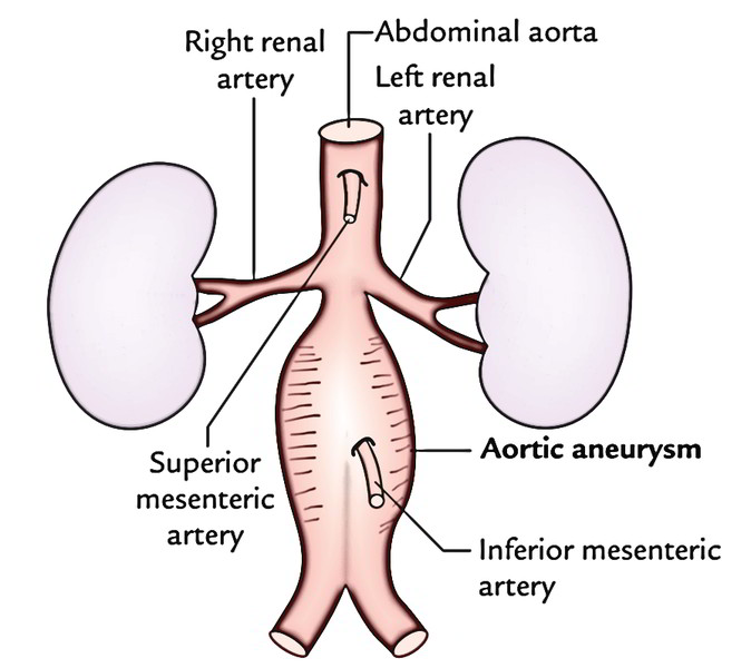
The aortic aneurysm (localized dilatation of the aorta) generally happens below the origin of the renal arteries (95%) typically in aged men. Most common cause of aortic aneurysm is atherosclerosis, which weakens the aortic wall. Medically, it presents as pulsatile and expansile abdominal mass normally just above and to the left of the umbilicus. The origin of renal arteries is a significant landmark in the abdominal aortic aneurysm surgery.

 (58 votes, average: 4.22 out of 5)