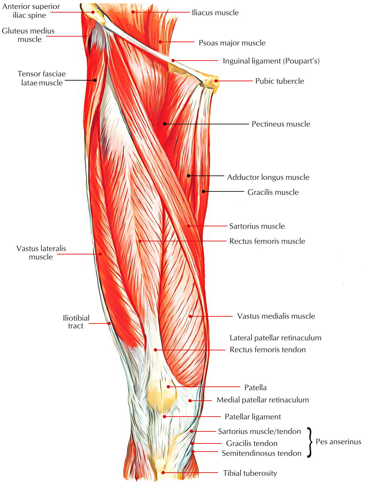
Anterior Compartment of the Thigh
Origin, Insertion, Nerve Supply, And Actions of The Muscles of The Front of The Thigh
| Muscle | Origin | Insertion | Nerve supply | Actions |
|---|---|---|---|---|
| 1. Quadriceps femoris (a) Rectus femoris | 1. Straight head: from the upper half of the anterior inferior iliac spine 2. Reflected head: from the groove above the acetabulum | Base of patella | Femoral nerve | Flexion of the hip joint and extension of the knee joint |
| (b) Vastus lateralis | 1. Upper part of the intertrochanteric line 2. Anterior and inferior borders of greater trochanter 3. Lateral lip of the gluteal tuberosity 4. Upper half of lateral lip of linea aspera | 1. Base and upper 1/3rd of the lateral border of the patella 2. Expansion to the capsule of the knee joint and iliotibial tract | Femoral nerve | Extension of the knee joint |
| (c) Vastus medialis | 1. Lower part of the intertrochanteric line 2 . Spiral line 3. Medial lip of the linea aspera 4.Upper 2/3rd of medial supracondylar line | 1. Base and upper 2/3rd of the medial border of the patella 2. Expansion to the capsule of the knee joint | Femoral nerve | Extension of the knee joint |
| (d) Vastus intermedius | Upper 3/4th of the anterior and lateral surfaces of the shaft of femur | Base of patella | Femoral nerve | Extension of the knee joint |
| 2. Articularis genu | Anterior surface of the lower part of the shaft of femur | Synovial membrane of the knee joint | Femoral nerve | Pulls up the synovial membrane of the knee joint during its extension |
| 3. Sartorius | Anterior superior iliac spine and upper half of the notch below it | Upper part of the medial surface of the shaft of the tibia | Femoral nerve | 1. Flexion of the hip and knee joints 2. Lateral rotation of the thigh |
| 4. Tensor fasciae latae | Outer lip of the iliac crest from anterior superior iliac spine to the tubercle of iliac crest | Iliotibial tract 3-5 cm below the greater trochanter | Superior gluteal nerve | Abduction of the hip joint |
Quadriceps Femoris
This muscle is thus referred to as because it is composed of 4 parts: rectus femoris, vastus lateralis, vastus medialis, and vastus intermedius. It’s supplied by the femoral nerve. It creates the majority of the mass on the anterior aspect of the thigh and is the strongest extensor of the knee joint.
All the parts of quadriceps cross only 1 joint, i.e., knee joint, with the exception of rectus femoris which crosses 2 joints, i.e., hip and knee joints.
Articularis Genu
It is composed of 3 or 4 muscular skids that are considered to be a separated part of the vastus intermedius.
Origin
It originates from the anterior surface of the lower part of the shaft of the femur, few centimeters above the patellar articular margin.
Insertion
Into the upper part of the synovial membrane of the knee joint.
Nerve Supply
It’s supplied by a twig from the nerve to vastus intermedius.
Actions
The articularis genu pulls up the synovial membrane upward to prevent its damage when the knee is extended.
Sartorius
It’s the longest muscle in the entire body, which crosses the front of the thigh obliquely from the lateral to the medial side.
Origin
It appears from the anterior superior iliac spine and upper half of the notch immediately below it.
Insertion
The muscle corkscrew obliquely through the thigh from lateral to medial side to reach the posterior aspect of the medial condyle of femur, where its tendon runs forwards to be added into the upper part of the medial outermost layer of the shaft of tibia in front of the insertions of gracilis and semitendinosus. The insertion of sartorius is inverted hockey stick -shaped.
Nerve Supply
It’s supplied by the anterior section of the femoral nerve.
Actions
It bends both the hip and the knee joints, and adducts and rotates the thigh laterally to bring the lower limb into the sitting position of a tailor (Latin sartor: tailor)/” palthi position” embraced by Indian Hindus during meal.
Tensor Fasciae Latae
It’s a short thick muscle, which is located at the junction of the gluteal region and the upper part of the front of the thigh.
Origin
It appears from the outer lip of the iliac crest extending from the anterior superior iliac spine to the tubercle of the crest.
Insertion
The muscle enters downward and somewhat backward and added into the iliotibial tract 3-5 cm below the level of the higher trochanter.
Nerve Supply
It’s supplied by the superior gluteal nerve.
Actions
It abducts the hip joint and keeps the extended position of the knee joint via the iliotibial tract.
Morphologically, the tensor fasciae latae is a muscle of the gluteal region that has migrated on the lateral aspect of the thigh during the course of development but keeps its Nerve Supply. It’s supplied by the superior gluteal nerve- the nerve of gluteal region.

 (50 votes, average: 4.71 out of 5)