📝 Feedback

Side of the Neck

It is about quadrangular in shape. It’s bounded superiorly by the lower border of the body of the mandible, anteriorly by anterior midline of the neck, posteriorly by anterior border of the trapezius and inferiorly by the clavicle and a line stretching from the angle of the mandible to the mastoid process.

Interactive Anatomical Interface


[HeadBones]

Highlight
Frontal
parietal
Temporal
Sphenoid
Zygomatic
Nasal
Lateral cartilage
greater alar cartilage
Mandbile
thyrohyoid membrane
cricotracheal ligament
cervical vertabrae
first rib
second rib

Highlight
Frontal
parietal
Temporal
Sphenoid
Zygomatic
maxilla
Nasal
Lateral cartilage
greater alar cartilage
Mandbile

Highlight
thyrohyoid membrane
thyroid cartilage
cricoid cartilage
cricotracheal ligament
ANterior longitudinal ligament
teeth
cervical vertabrae
first rib
second rib
[HeadBonesSide]

Highlight
frontal
Parietal
Sphenoid
temporal
Zygomatic
Maxilla
Lateral cartilage
posterior atlantoaxial ligament
lateral ligament
greater alar cartilage
teeth
mandible
hyoid
first rib
posterior atlanto-occipital memebrane
thyrohyoid membrane
thyroid cartilage
ricothyroid membrane and ligament
cricoid cartilage
cricotracheal ligament
t1 vertebra
joint capsule
Anterior longitudinal ligamnent
nuchal ligament
Cervical Vertebra
lacrimal

Highlight
frontal
Parietal
Sphenoid
temporal
Zygomatic
Maxilla
Lateral cartilage
greater alar cartilage
mandible
hyoid
thyrohyoid membrane
thyroid cartilage
ricothyroid membrane and ligament
cricoid cartilage
lateral ligament
cricotracheal ligament

Highlight
stylohyoid ligament
Anterior longitudinal ligamnent
nuchal ligament
Cervical Vertebra
first rib
t1 vertebra
lacrimal
occipital bone
teeth
posterior atlanto-occipital memebrane
posterior atlantoaxial ligament
joint capsule
stylomandibular ligament

[InnerSkull]

Highlight
nasal
occipital bone
Internal auditory meatus
Stylomastoid Foramen“>Stylomastoid foramen
Temporal
sphenoid
ethmoid
zygomatic bone
perpendicular plate
lacrimal bone
nasal cartilage
vomer“>vomer
Nasal concha
Mandible
tectorial membrane of atlanto-axial joint
Foramen magnum“>foramen magnum
tectorial membrane
Crista Galli“>crista galli
ethmoidal sinuses
cribriform plate“>cribriform plate
Foramen rotundum“>foramen rotundum
optic foramen
Superior orbital fissure“>superior orbital fissure
palatine
nuchal ligament
c01 atlas
posterior atlantoaxial ligament
c03 vertebrae
c04 vertebrae
C5 vertebrae

Highlight
nasal
temporal bone
occipital bone
Stylomastoid foramen
Temporal
sphenoid
ethmoid
zygomatic bone
zygomatic bone
lacrimal bone
perpendicular plate
posterior atlantoaxial ligament
nasal cartilage
vomer
Nasal concha
palatine
tectorial membrane of atlanto-axial joint

Highlight
foramen magnum
tectorial membrane
c1 atlas
crista galli
lacrimal bone
Mandible
ethmoidal sinuses
cribriform plate
foramen rotundum
Internal auditory meatus
optic foramen
superior orbital fissure
nuchal ligament
c01 atlas
c2 axis
c03 vertebrae
c04 vertebrae
C5 vertebrae

Please hover over the figure to get a brief info about the body parts.
To get the elaborated info click on the body part.


Please rotate the device for better visual interaction.
 

Like lymphadenopathy, cysts, tumors, etc, the side of the neck is the common site for different pathological lesions. For performing different clinical procedures like biopsy, venepuncture, nerve block, etc it is also a regular site.

Surface Landmarks on the Side of the Neck

The most significant landmark on the side of the neck is sternocleidomastoid type. It is viewed as a raised ridge extending obliquely from the sternum to the mastoid process and becomes notable when the face is turned to the opposite side.

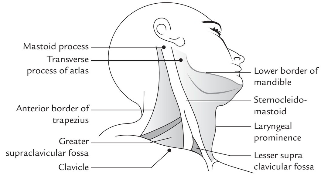
  • Mastoid process is a large bony projection easily felt behind the lower part of the auricle.
  • Anterior border of trapezius becomes notable when shoulder is elevated against the resistance.
  • Lesser supraclavicular fossa is a small triangular depression above the medial end of the clavicle between the sternal and clavicular heads of sternocleidomastoid. It overlies the internal jugular vein.
  • Greater supraclavicular fossa is a depression above the middle one-third of the clavicle. It overlies the cervical part of brachial plexus and the third part of the subclavian artery.
  • Transverse process of first cervical vertebra can be felt on deep pressure midway between the angle of the mandible and mastoid process. It’s crossed by spinal accessory nerve.
  • Lower border of the mandible can be easily felt by running a finger backwards from chin to the angle of the mandible.
  • Clavicle being subcutaneous can be felt along its whole extent in the junction of the neck and chest.

Rate this Article: 1 Star2 Stars3 Stars4 Stars5 Stars (51 votes, average: 4.59 out of 5)
Loading...
x
Trusted By The World’s Best