The muscles in the submandibular region create 4 muscular planes; from superficial to deep, from the perspective of surgical procedures these are:
- First muscular plane: created by the digastric and stylohyoid muscles
- Second muscular plane: created by the mylohyoid muscle
- Third muscular plane: created by the geniohyoid, hyoglossus and styloglossus muscles
- Fourth muscular plane: created by the genioglossus and a part of superior constrictor of the pharynx
Muscles of First Muscular Plane
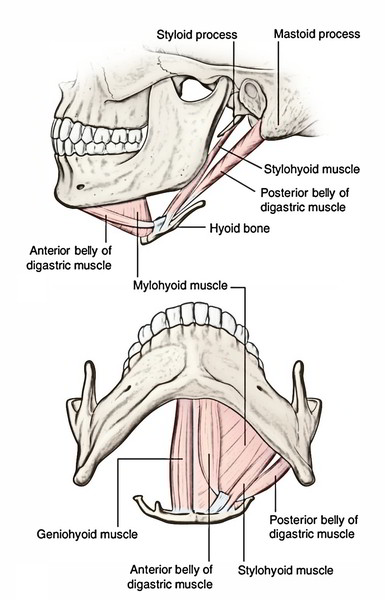
Digastric Muscle
It is made up of posterior and anterior bellies and is a strap like muscle, united by an intermediate tendon. This muscle is so called because it has 2 abdomens.
Origin
From the mastoid notch of the temporal bone, the bipinnate posterior abdomen originates. From the digastric fossa on the lower border of the mandible near the symphysis ment, the unipinnate anterior abdomen originates.
Insertion
Posterior abdomen enters downwards and forwards between the carotid triangle below and behind and digastric triangle above and in front and fit into the intermediate tendon.
Anterior abdomen enters downwards and backwards on the mylohyoid to be added into the intermediate tendon.
The intermediate tendon is anchored to the junction of the body and greater cornu of hyoid bone by an inverted U-shaped facial sling of investing layer of deep cervical fascia. The tendon of digastric muscle enters between the 2 slips of tendon of stylohyoid muscle.
Nerve Supply
The posterior abdomen grows from mesoderm of the second pharyngeal arch and thus, supplied by the facial nerve.
The anterior abdomen grows from the first pharyngeal arch and, thus, supplied by the mylohyoid nerve, a branch of inferior alveolar nerve from mandibular nerve.
Activities
- Helps to depress the mandible when the mouth is opened extensively against resistance.
- Pulls the hyoid bone upwards during deglutition.
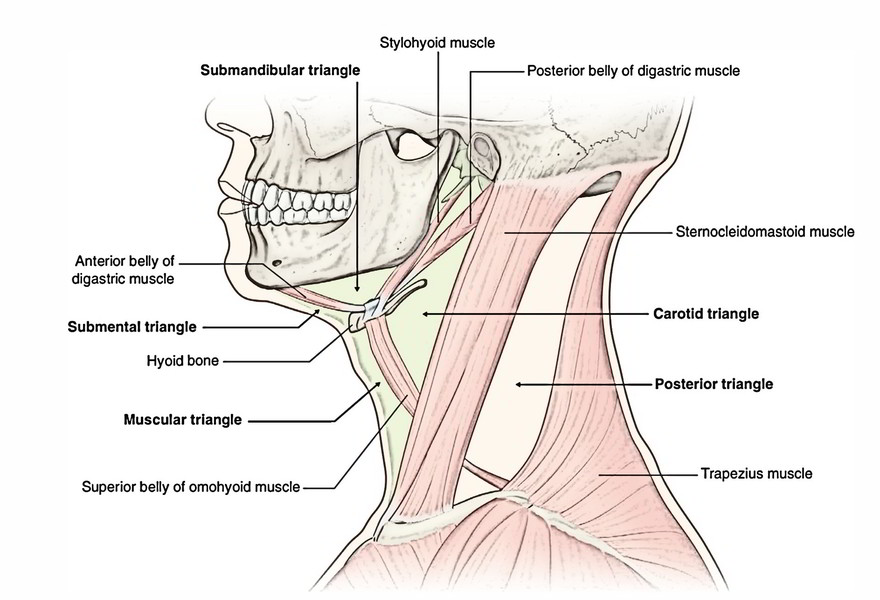
Connections of the Posterior Belly of the Digastric Muscle
Superficial:
- Skin, superficial fascia, platysma and investing layer of deep cervical fascia.
- Mastoid process and sternocleidomastoid muscle.
- Parotid gland and the angle of the mandible.
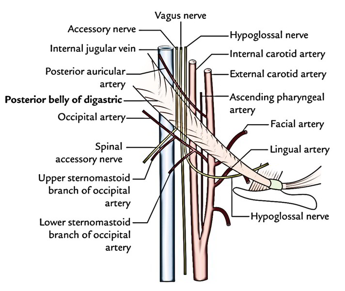
Deep:
Neurovascular bundle of neck composed of internal jugular vein, external and internal carotid arteries 10th, 11th and 12th cranial nerves.
Upper border: Stylohyoid muscle and posterior auricular artery run along the upper border of the posterior belly of the digastric.
Lower border: Occipital artery runs along and under the cover of the lower border of digastric.
The relationships of posterior belly of digastric are essential because 3 cranial nerves: 10th, 11th and 12th and 3 great blood vessels of neck, viz. internal jugular vein, internal and external carotid arteries pass deep to it.
Differences between the Anterior and Posterior Belly of the Digastric Muscle
| Anterior belly | Posterior belly |
|---|---|
| Unipennate | Bipennate |
| Develops from 1st pharyngeal arch | Develops from 2nd pharyngeal arch |
| Supplied by mylohyoid nerve (nerve of 1st pharyngeal arch) | Supplied by facial nerve (nerve of 2nd pharyngeal arch) |
Stylohyoid Muscle
It’s a slight muscle that is located along the upper border of the posterior belly of the digastric muscle.
Origin
It originates from the posterior surface of the styloid process.
Insertion
It’s fit into the hyoid bone in the junction between the body and greater cornu. At the insertion, its tendon divides into 2 slides that pass 1 on either side of the intermediate tendon of the digastric muscle.
Nerve Supply
The stylohyoid muscle grows from the second arch and, thus, it’s furnished by the facial nerve.
Activities
It pulls the hyoid bone upwards and backwards and elongates the floor of the mouth.
The stylohyoid muscle is thought to be the delaminated portion of the posterior belly of the digastric muscle.
Muscles of Second Muscular Plane
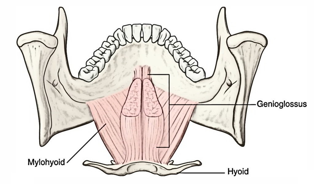
Mylohyoid Muscle
It’s a flat, triangular muscle being located deep to the anterior belly of the digastric muscle. The left and right mylohyoid muscles join in the median fibrous raphe to create the gutter-shaped floor of the mouth; over which is located the tongue, therefore the floor of the mouth is also termed diaphragma oris.
Origin
From the mylohyoid line of the mandible.
Insertion
The fibres run downwards and medially. The posterior fibres are added into the body of the hyoid bone. The middle and anterior fibres are added into the median fibrous raphe stretching from symphysis menti to the hyoid bone.
Nerve Supply
The mylohyoid muscle grows from the very first pharyngeal arch, consequently it’s supplied by mylohyoid nerve, a branch of inferior alveolar nerve from mandibular nerve.
Activities
The mylohyoid muscle elevates the floor of the mouth and for this reason the tongue during the very first phase of the deglutition.
It also helps in the depression of the mandible against resistance.
It repairs or elevates the hyoid bone.
Muscles of Third Muscular Plane
Geniohyoid Muscle
It’s a narrow muscle that is located alongside the midline deep to the mylohyoid.
Origin
From inferior genial tubercle of the mandible.
Insertion
The fibres run backwards and downwards to be added into the anterior surface (front) of the body of the hyoid bone, above the medial part of the mylohyoid muscle.
Nerve Supply
C1 fibres via hypoglossal nerve.
Activities
- The geniohyoid elevates the hyoid bone by pulling it upwards and forwards and thereby shortens the floor of the mouth.
- They may depress the mandible when the hyoid bone is fixed.
Hyoglossus Muscle
It’s the flat quadrilateral muscle of the tongue.
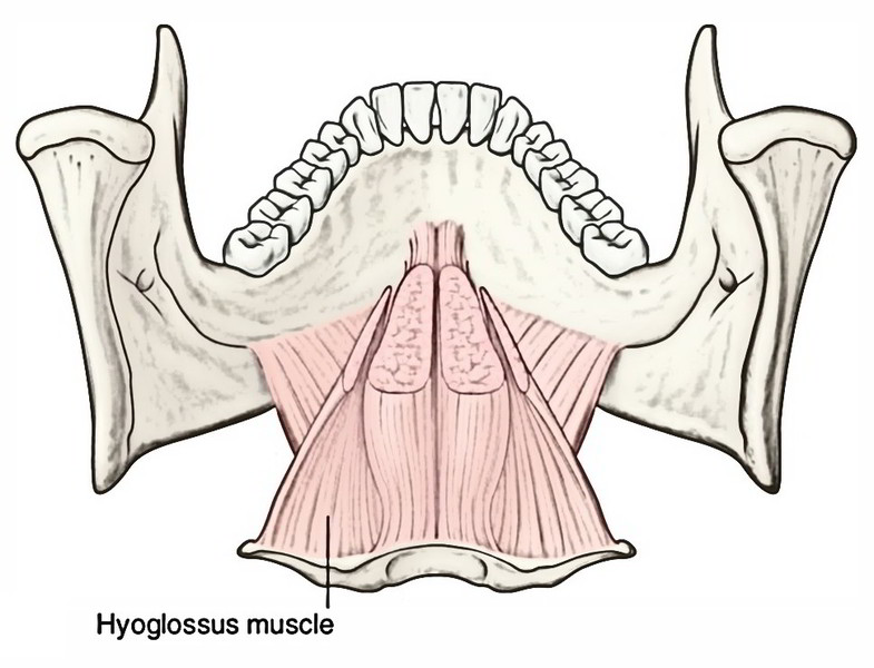
Origin
From upper surface of the entire length of the greater cornu and adjacent part of the body of hyoid bone.
Insertion
Into the side of tongue between styloglossus laterally and inferior longitudinal medially. The fibres of hyoglossus from hyoid bone run upwards and somewhat forwards and decussate with the fibres of styloglossus.
A part of hyoglossus might be connected to the lesser cornu of the hyoid bone and create a different muscle referred to as chondroglossus.
Connections
The hyoglossus, a quadrilateral sheet of muscle, is the key muscle of the suprahyoid region because it acts as a landmark for contiguous structures in the region. For that reason, its connections are extremely significant to surgeons.
Superficial relationships:
The superficial connections of the hyoglossus are as follows:
- Hypoglossal nerve, crosses the lower part of muscle from behind forwards.
- Lingual nerve, crosses the upper part of muscle from behind forwards.
Deep part of the submandibular gland and submandibular duct:
The gland is located in the middle of hyoglossus muscle and the duct is located between the gland and the muscle.
Submandibular ganglion is located between the lingual nerve and deep part of the submandibular gland
- Styloglossus muscle- interdigitates with hyoglossus.
Mylohyoid muscle overlaps the hyoglossusanterosuperiorly.
Deep connections:
The deep relationships of the hyoglossus are as follows:
- Inferior longitudinal muscle of tongue.
- Genioglossus muscle anteriorly.
- Middle constrictor of pharynx posteriorly.
- Glossopharyngeal nerve.
- Stylohyoid ligament.
- Lingual artery.
Structures Passing Deep To Posterior Border of Hyoglossus
From above downwards, these are as follows:
- Glossopharyngeal nerve.
- Stylohyoid ligament.
- Lingual artery.
Nerve Supply
The hyoglossus muscle grows from occipital myotomes, for that reason, it’s supplied by hypoglossal nerve.
Activities
- Depresses the side of tongue to make the dorsal surface of the tongue convex.
- Helps in the retraction of the protruded tongue.
Styloglossus Muscle
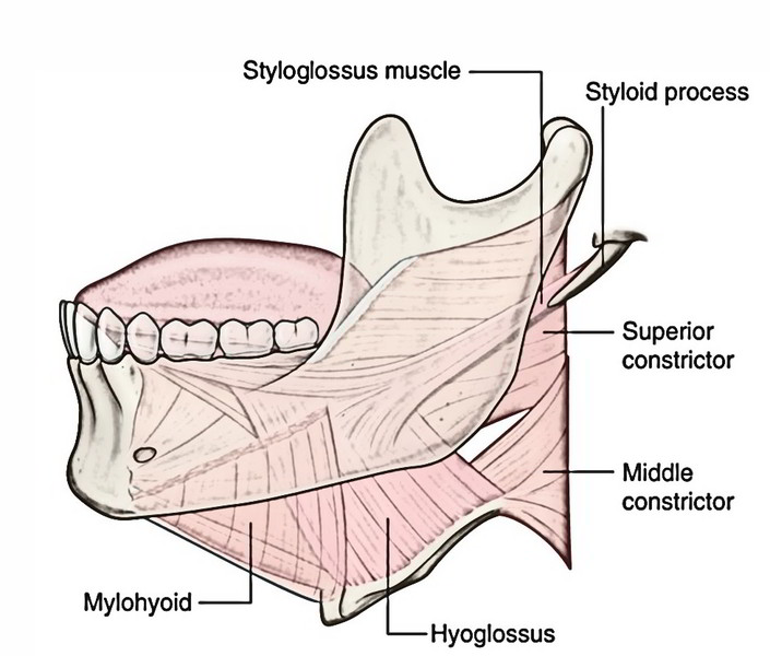
Origin
From front of the tip of styloid process and adjoining part of stylohyoid ligament.
Insertion
The fibres run forwards to be added into the full length of the side of tongue, interdigitating with the fibres of the hyoglossus muscle.
Nerve Supply
It’s supplied by hypoglossal nerve.
Activities
Retracts the tongue upwards and backwards and consequently it’s antagonist to the genioglossus. Together with its counterpart of opposite side, it creates a median gutter on the dorsum of tongue for the passage of food.
Muscles of Fourth Muscular Plane
Genioglossus Muscle
It’s a fan-shaped extrinsic muscle of tongue and together with its counterpart of the opposite side creates the majority of the majority of the tongue.
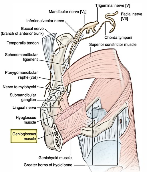
Origin
From superior genial tubercle of the mandible.
Insertion
- The fibres radiate backwards fan-wise into the substance of the corresponding half of the tongue alongside the median septum, from the tip to the base, for insertion.
- Lower fibres are added into the body of the hyoid and create the root of the tongue.
- Intermediate fibres pass underneath the anterior border of the hyoglossus and go backwards up to stylohyoid ligament and middle constrictor of the pharynx.
- Upper fibres turn upwards and forwards to go up to the tip of the tongue.
Nerve Supply
It’s supplied by hypoglossal nerve.
Activities
The muscles of both sides together protrude the tongue and make an elongated gutter on the dorsal surface of the tongue for the passage of food.
Test Your Knowledge
Muscles of the Submandibular Region

 (50 votes, average: 4.70 out of 5)