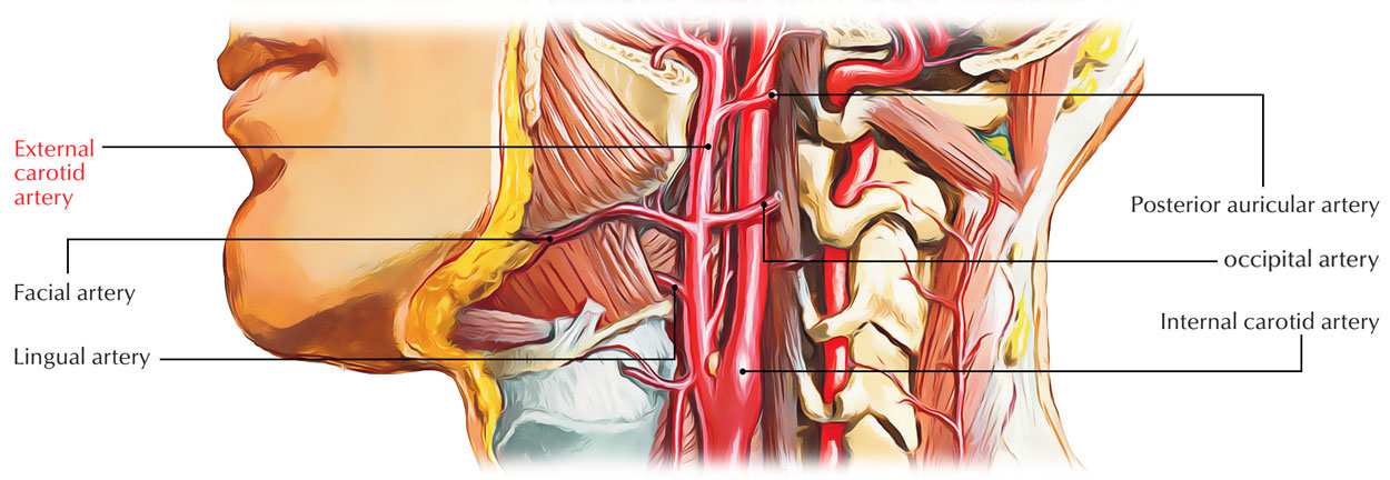
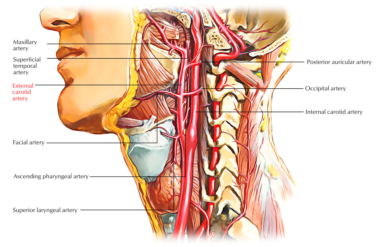
Origin
The external carotid arteries emerge at the level of the upper margin of the thyroid cartilage which is located at the plane of the fourth cervical vertebra. It takes a slightly arced path upwards as well as anteriorly before inclining back towards the space posterior to the neck of the mandible.

External Carotid Arteries
Insertion
Generally, the external carotid artery is located anterior towards the internal carotid artery as it rises upwards within the carotid triangle. It goes posterior towards the posterior belly of the digastric inside the retromandibular fossa.
The anterior branches of the external carotid overlap the internal carotid in some of cases where the internal carotid artery is anteromedial towards the external carotid artery. This helps in distinguishing the external carotid artery from the internal carotid artery.
Branches

Branches of External Carotid Arteries
The external carotid arteries begin to produce branches directly after the division of the common carotid arteries:
Superior thyroid artery
It originates just beneath the tip of the greater cornu of the hyoid bone from the front of external carotid artery. It travels downwards and forwards, parallel and superficial towards the external laryngeal nerve in order to connect with the upper end of the thyroid gland, which is supplied by it.
Branches
(a) Infrahyoid branch anastomoses with its corresponding one of opposite side.
(b) Sternomastoid branch towards the sternomastoid muscle.
(c) Superior laryngeal artery goes alongside the internal laryngeal nerve as well as perforates the thyrohyoid membrane in order to circulate the larynx while it travels deep towards the thyrohyoid muscle.
(d) Cricothyroid branch travels across the cricothyroid ligament and anastomoses with its corresponding branch of the contrasting side.
(e) Glandular branches towards the thyroid gland. One of the branches anastomoses along the upper border of the isthmus of the thyroid gland with its equivalent of the opposite side.
Lingual artery
It originates from the front of the external carotid artery opposed to the tip of the greater cornu of the hyoid bone. It is the chief artery to supply blood towards the tongue. It may arise along the facial artery. By the hyoglossus muscle it is separated into three parts:
(a) First part is located in the carotid triangle and creates a distinctive loop with convexity upwards over the greater cornu. Superficially, the loop is traversed by the hypoglossal nerve. The loop enables free movement of the hyoid bone.
(b) Second part along the upper border of the hyoid bone is located deep towards the hyoglossus muscle.
(c) Third part a.k.a. deep lingual artery or arteria profunda linguae first travels upwards alongside the anterior margin of the hyoglossus muscle and then forward on the undersurface of the tongue, where it anastomoses with its fellow of opposite side.
Branches
(a) First part— suprahyoid branch that anastomoses with its fellow of opposite side.
(b) Second part— generally two dorsal linguae branches towards the dorsum of tongue along with tonsil.
(c) Third part—sublingual artery towards the sublingual gland.
Facial artery (a.k.a. external maxillary artery)
It originates from the front of the external carotid artery just above the tip of the greater cornu of the hyoid bone.
Parts
(a) Cervical part of the facial artery rises deep towards the digastric as well as stylohyoid muscles, travels where it channels the posterior border of the submandibular gland, deep towards the ramus of mandible. Afterwards it creates S-shaped turn, first over the submandibular gland curving down with convexity upwards and then above the base of the mandible upwards with convexity downwards.
(b) Facial part of the facial artery emerges nearby the lower margin of the body of the mandible where the facial artery coils at the anteroinferior angle of the masseter.
Branches
From the cervical part – branches in the neck:
(a) Ascending palatine artery originates near the origin of facial artery, ascends, as well as accompanies the levator palati, travels over the upper margin of the superior constrictor as well as circulates primarily the palate.
(b) Tonsillar artery a.k.a. main artery of tonsil perforates the superior constrictor and terminates within the tonsil.
(c) Glandular branches circulate the submandibular gland.
(d) Submental artery travels forwards on the mylohyoid muscle along mylohyoid nerve.
It supplies:
- Mylohyoid muscle.
- Submandibular and sublingual salivary glands.
Occipital artery
It originates from the posterior part of the external carotid artery on the same plane as the facial artery.
- It travels backwards and upwards beneath cover of lower margin of the posterior belly of digastric muscle superficial towards crosses the apex of the posterior triangle, along with internal carotid artery, internal jugular vein, and last four cranial nerves.
- Then it goes deep towards the mastoid process turning over the lower surface of the temporal bone medial towards the mastoid notch.
Branches
(a) Sternomastoid branches are generally two in total. They travel downwards and backwards, and supply the sternocleidomastoid. The upper one goes together with the spinal accessory nerve and lower one is curved by the hypoglossal nerve.
(b) Mastoid branch goes inside the cranial cavity via mastoid foramen and circulates mastoid air cells.
(c) Meningeal branches go inside the cranial cavity via jugular foramen and hypoglossal canal in order to supply dura mater of posterior cranial fossa.
(d) Muscular branches circulate adjoining muscles.
(e) Auricular branch supply the cranial surface of the auricle.
(f) Descending branch splits into superficial as well as deep branches:
- Superficial branch anastomoses with the superficial branch of transverse cervical artery.
- Deep branch anastomoses with the deep cervical artery.
Posterior auricular artery
It originates a little above the occipital artery from the posterior aspect of the external carotid artery. It crosses superficial to the stylohyoid muscle. Along the upper border of the posterior belly of digastric muscle and deep towards the parotid gland, it travels upwards and backwards parallel towards the occipital artery. Afterwards it becomes superficial and is located behind the ear which it supplies on the base of mastoid process.
Branches
(a) Stylomastoid artery enters the stylomastoid foramen in order to supply middle ear, mastoid air cells, along with facial nerve.
(b) Auricular branch supplies both cranial as well as lateral surfaces of the auricle.
(c) Occipital branch supplies scalp above as well as behind the auricle.
Ascending pharyngeal artery
It originates from the medial part of the external carotid artery near its lower end. It runs in the middle of the side wall of the pharynx as well as internal carotid artery, vertically upwards till the base of the skull.
Branches
(a) Pharyngeal and prevertebral branches to corresponding muscles.
(b) Meningeal branches, which traverse foramina in the base of the skull.
(c) Inferior tympanic supplies medial wall of tympanic cavity.
(d) Palatine branches go with levator veli palatini towards the palate.
Superficial temporal artery
- It is the smaller but more direct concluding branch of the external carotid artery.
- It arises behind the neck of the mandible deep towards the upper portion of the parotid gland.
- It travels vertically upwards passing the root of zygoma in front of the tragus.
- Above the zygoma, it divides into anterior and posterior branches that circulate the temple and scalp.
Branches
- Transverse facial artery runs forwards through the masseter beneath the zygomatic arch.
- Anterior auricular branch circulates the lateral surface of auricle and external auditory meatus.
- Zygomatico-orbital artery runs forwards along the upper border of zygomatic arch in the middle of two layers of temporal fascia and touches the lateral angle of the eye.
- Middle (deep) temporal artery runs on the temporal fossa deep to temporalis muscle and supplies temporalis muscle and fascia.
- Anterior and posterior terminal branches.
- The anterior branch supplies the muscles and skin of the frontal region.
- The posterior branch supplies skin and the auricular muscles.
Maxillary Artery
The maxillary artery originates posterior towards the neck of the mandible, it travels from the parotid gland, proceeds medial towards the neck of the mandible and inside the infratemporal fossa, and carries on from this zone into the pterygopalatine fossa.
Relations
The external carotid artery is enclosed by:
- Skin
- Superficial fascia
- Platysma
- Deep fascia
- Anterior margin of the Sternocleidomastoideus muscle
It is crossed by:
- Hypoglossal nerve.
- The lingual, ranine, common facial and superior thyroid veins.
- Digastricus and Stylohyoideus muscles.
Higher up it travels deeply inside the substance of the parotid gland, where it is located deep towards the facial nerve and the joint of the temporal and internal maxillary veins.
- Medial to it are
- Hyoid bone,
- Wall of the pharynx,
- Superior laryngeal nerve
- A portion of the parotid gland
- Lateral to it, in the lower part of its course, is the internal carotid artery.
- Posterior towards it, near its beginning, is the superior laryngeal nerve; and higher up, from the internal carotid it is divided through:
- Styloglossus and Stylopharyngeus muscles
- Glossopharyngeal nerve
- Pharyngeal branch of the vagus
- Part of the parotid gland
Functions
Branches of external carotid artery supply:
- Superior thyroid artery
- Thyrohyoid muscle
- Internal structures of the larynx
- Sternocleidomastoid and cricothyroid muscles
- Thyroid gland
- Ascending pharyngeal artery
- Pharyngeal constrictors and stylopharyngeus muscle
- Palate
- Tonsil
- Pharyngotympanic tube
- Meninges in posterior cranial fossa
- Lingual artery
- Muscles of the Tongue
- Palatine Tonsil
- Soft Palate
- Epiglottis
- Floor of Mouth
- Sublingual Gland
- Facial artery
- All structures in the face from the inferior border of the mandible anterior to the masseter muscle towards the medial corner of the eye, the soft palate, palatine tonsil, pharyngotympanic tube, submandibular gland
- Occipital artery
- Sternocleidomastoid Muscle
- Meninges in Posterior Cranial Fossa
- Mastoid Cells
- Deep Muscles of the Back
- Posterior Scalp
- Posterior auricular artery
- Parotid Gland and Nearby Muscles
- External Ear and Scalp Posterior to Ear
- Middle and Inner Ear Structure
- Superficial temporal artery
- Parotid Gland and Duct
- Masseter Muscle
- Lateral Face
- Anterior Part of External Ear
- Temporalis Muscle
- Parietal and Temporal Fossae
- Maxillary artery
- External acoustic meatus
- Lateral and medial surface of tympanic membrane
- Temporomandibular joint
- Dura mater on lateral wall of skull and inner table of cranial bones
- Trigeminal ganglion and dura in vicinity
- Mylohyoid muscle
- Mandibular teeth
- Skin on chin
- Temporalis muscle
- Outer table of bones of skull in temporal fossa
- Structures in infratem poral fossa
- Maxillary sinus
- Upper teeth and gingivae
- Infra-orbital skin
- Palate
- Roof of pharynx
- Nasal cavity

 (60 votes, average: 4.73 out of 5)