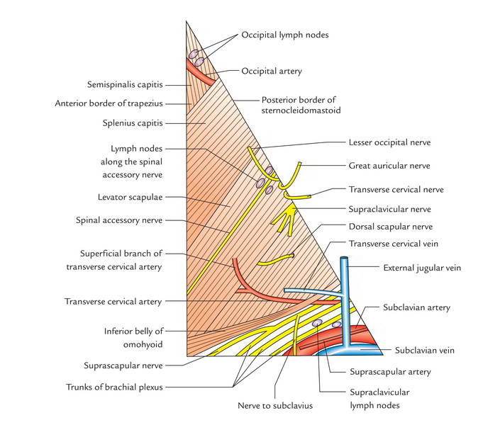
Behind the sternocleidomastoid muscle, the triangular space on the side of neck is called the Posterior triangle. Its base downwards in the direction of the clavicle and apex is pointed upwards and backwards in the direction of the mastoid process.
Borders
- Anterior: Posterior border of sternocleidomastoid muscle.
- Posterior: Anterior border of trapezius muscle.
- Inferior (base): Superior aspect of middle third of the clavicle.
- Superior (apex): Meeting point of sternocleidomastoid and trapezius muscles at the superior nuchal line of the occipital bone.
Roof
It’s created by the investing layer of the deep cervical fascia, extending between the sternocleidomastoid and trapezius muscles.
The superficial fascia overlying the roof includes platysma, external jugular and posterior jugular veins and cutaneous nerves and vessels.
Structures piercing the roof of the posterior triangle are:
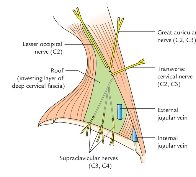
4 cutaneous branches of cervical plexus, viz.
- Lesser occipital nerve (C2).
- Great auricular nerve (C2, C3).
- Transverse cervical nerve (C2, C3).
- Supraclavicular nerves (C3, C4).
They pierce the roof near the middle of the posterior border of the sternocleidomastoid muscle.
External jugular vein: It starts just below the angle of mandible, runs downwards and backwards crossing the sternocleidomastoid obliquely and under the cover of platysma.
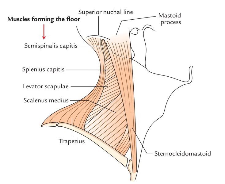
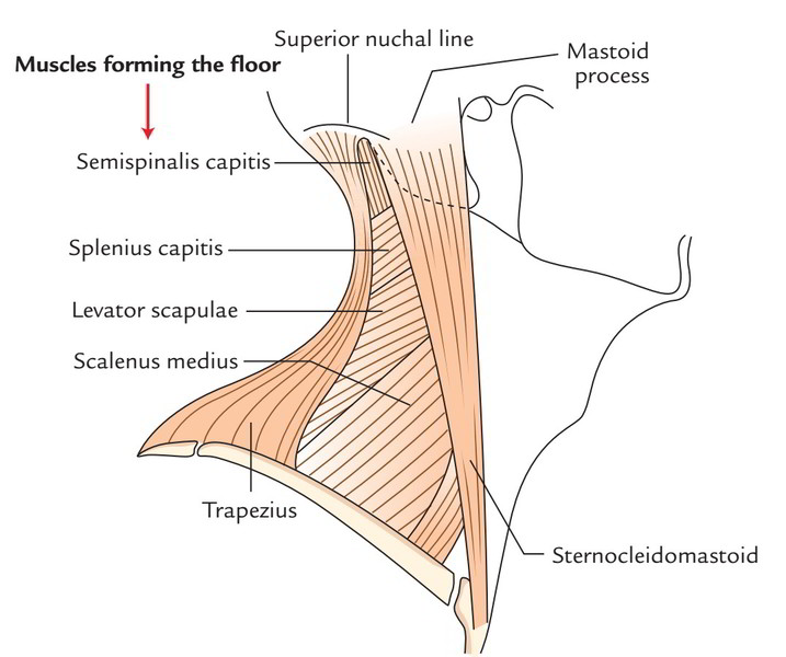
Floor
The floor of posterior triangle is muscular and is composed from above downwards by these muscles:
- Semispinalis capitis.
- Splenius capitis.
- Levator scapulae.
- Scalenus medius.
- First digitation of serratus anterior (occasionally).
Fascial Carpeting of the Posterior Triangle
The muscular floor of posterior triangle is covered by prevertebral layer of deep cervical fascia, which creates the fascial carpeting of the floor of the posterior triangle. It creates axillary sheath around subclavian artery and brachial plexus going from the root of the neck to the upper limb.
The lower part of the posterior triangle is crossed by inferior belly of omohyoid superficial to the fascial carpeting.
Clinical Significance
Pus gathered in the posterior triangle deep to its fascial carpeting from tubercular cervical vertebrae may track downwards and laterally along the axillary sheath to first appear in the axilla or even in the arm afterwards.
Subdivisions
The posterior triangle is subdivided into 2 parts by the inferior belly of the omohyoid, which crosses the lower part of the triangle obliquely upwards and forwards (a) a bigger upper part named occipital triangle and (b) a small lower part termed subclavian (supraclavicular) triangle. These parts are thus termed since they include occipital and subclavian arteries, respectively.
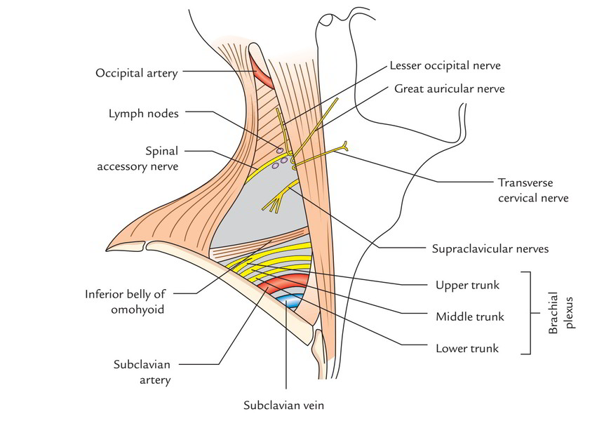
Contents

- In the occipital triangle (i.e., above the omohyoid).
- Spinal accessory nerve.
- third and 4th cervical nerves supplying branches to levator scapulae and trapezius muscles.
- Dorsal scapular nerve (C5).
- 4 cutaneous branches of cervical plexus (first parts).
- Superficial transverse cervical artery.
- Occipital artery.
- In the subclavian/supraclavicular triangle (i.e., below the omohyoid).
- third part of the subclavian artery.
- Subclavian vein.
- Terminal part of external jugular vein.
- Trunks of brachial plexus.
- Superficial (transverse) cervical, suprascapular and dorsal scapular arteries.
- Lymph nodes.
Key Points
The most essential contents of posterior triangle are:
(a) Third part of subclavian artery
(b) Brachial plexus (cervical part)
(c) Spinal accessory nerve and
(d) Lymph nodes.
All the significant contents of the posterior triangle are located deep to the fascial carpeting of the floor with the exception of spinal accessory nerve, which is located just underneath the roofing. In procedures on the posterior triangle all the structures with the exception of spinal accessory nerve are safe, supplied fascial carpeting of posterior triangle is left undamaged.
Important Features of Some of the Contents
Spinal accessory nerve
This nerve comes in the posterior triangle by piercing the posterior border of the sternocleidomastoid (a little above the middle of the border). In this case, it’s related to lymph nodes of the upper deep cervical chain. The nerve then crosses the posterior triangle by running downwards and laterally around and parallel to the fibres of levator scapulae muscle to evaporate below to the anterior border of trapezius (about 5-6 cm above the clavicle) and supplies trapezius muscle. In the posterior triangle it is adherent to the deep aspect of the fascial roof of the triangle.
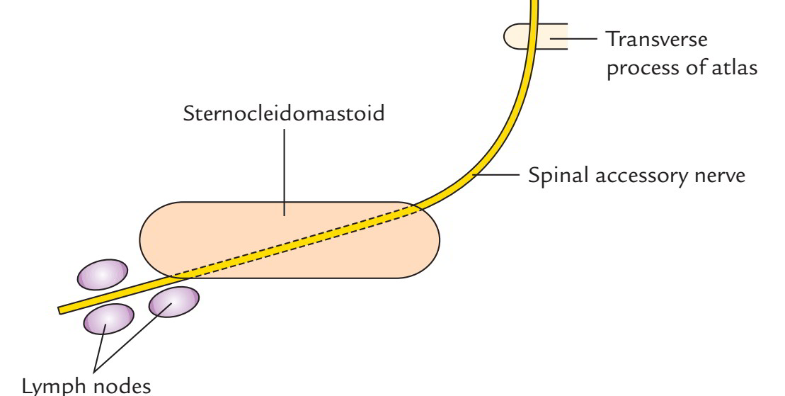
More proximally, the nerve is located in front of transverse process of atlas vertebra runs downwards and laterally to goes into the anterior border of sternocleidomastoid muscle between its upper 2 quarters and supplies it.
Clinical significance
The spinal accessory nerve could be damaged in operations requiring the removal or biopsy of lymph nodes in the posterior triangle of the neck.
4 cutaneous branches of cervical plexus
In spite of the fact that cervical plexus lies deep to the sternocleidomastoid but its cutaneous branches come in the midpoint or just above the midpoint of the posterior border of the sternocleidomastoid by piercing the deep cervical fascia.
The course and distribution of these cutaneous nerves are as follows:
- The lesser occipital nerve hooks around the spinal accessory nerve and ascends for a short space along the posterior border of sternocleidomastoid to inner-vate the skin of the upper one-third of the cranial outermost layer of the auricle and that of the head supporting the auricle.
- The great auricular nerve runs forwards and upwards across the sternocleidomastoid in the direction of the angle of mandible where it divides into anterior and posterior branches. It first supplies the skin of the face over the angle of the mandible and after that the skin over the mastoid region and lower part of both the surfaces of the auricle.
- The transverse cervical nerve enters forward across the sternocleidomastoid deep to the external jugular vein and after that splits into ascending and descending branches to supply the skin of the front of the neck.
- The supraclavicular nerve appears as a common trunk which descends downwards and splits into medial, intermediate and lateral supraclavicular nerves.
- The medial supraclavicular nerve crosses in front of the medial one-third of the clavicle to supply the skin on the chest up to the second rib.
- The intermediate supraclavicular nerve enters in front of the middle third of the clavicle to supply the skin on the very front of the chest. Sometimes it pierces the clavicle via and through.
- The lateral supraclavicular nerve crosses in front of the lateral third of the clavicle and supplies the skin over the shoulder and the upper half of the deltoid muscle.
- The point in the junction of the upper and middle third of the posterior border of sternocleidomastoid where 4 cutaneous nerves and spinal accessory nerve issue is referred to as nerve point of the neck.
- In ‘cervical plexus nerve block’ the anesthetic agent is injected at this site.
- Muscular branches to levator scapulae and trapezius (C3, C4).
- They appear at concerning the middle of sternocleidomastoid. The branches going to levator scapulae shortly finish in it, on the other hand those going to trapezius run below and parallel to the spinal accessory nerve and cross the middle of the triangle.
Nerve to rhomboids:
- It pierces scalenus medius muscle and instantaneously enters deep to levator scapulae muscle.
- Cervical part of the brachial plexus.
- The roots and trunks of brachial plexus are located in the neck.
- 4 branches originate from cervical part of brachialplexus.
- 4 branches originate from cervical part of brachialplexus- 2 from roots and 2 from trunks.
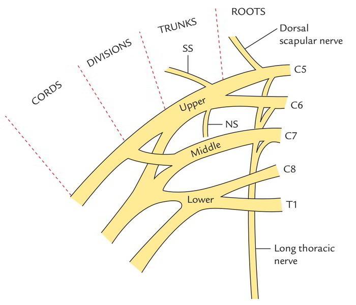
From roots:
Dorsal scapular nerve originates from C5 root. It pierces the scalenus medius and runs laterally across it to pass deep to levator scapulae, which it furnishes.
Long thoracic nerve (or nerve to serratus anterior) originates from C5, C6 and C7 roots. It enters downwards behind the brachial plexus and third part of the subclavian artery. It crosses the first rib to supply the serratus anterior muscle.
Remember that C5 and C6 roots pierce the scalenus medius and C7 root joins the nerve at the lower level in the axilla.
From trunks:
Nerve to subclavius originates from upper trunk (C5 and C6). It enters downwards in front of brachial plexus and subclavian vessels to supply the subclavius muscle.
Suprascapular nerve also originates from the upper trunk (C5 and C6). It enters laterally deep to inferior belly of omohyoid and trapezius to goes into the supraspinous fossa via suprascapular notch to supply the supraspinous and infraspinatus muscles.
Transverse cervical artery:
It’s a branch of thyrocervical trunk of the initial part of subclavian artery. It enters laterally and upwards crossing the scalenus anterior, upper trunk of brachial plexus and scalenus medius.
At the lower border of levator scapulae, it splits into the superficial and deep branches.
Suprascapular artery: This is also a branch of thyrocervical trunk. It enters laterally and backwards behind the clavicle to get to the upper border of the scapula.
Dorsal scapular artery: It originates from the third part of the subclavian artery. It enters laterally in front of scalenus medius and via the brachial plexus to get to the superior angle of the scapula.
Occipital artery: It’s a branch of external carotid artery. It crosses the apex of the triangle superficial to the semispinalis capitis.
Subclavian artery: It enters behind the scalenus anterior over the very first rib. It’s closely associated with the lower trunk of the brachial plexus.
Subclavian vein: It enters in front of scalenus anterior over the very first rib.
Terminal part of external jugular vein: It pierces the fascial roof of the posterior triangle about 2.5 cm above the clavicle to terminate in the subclavian vein.
Lymph nodes: These are deep cervical lymph nodes seen at these sites in the posterior triangle:
- A chain of nodes along the posterior border of sternocleidomastoid.
- A chain of nodes along the spinal accessory nerve.
- A few nodes in the apical region of the triangle termed occipital lymph nodes.
- A group of lymph nodes in the supraclavicular part of triangle referred to as supraclavicular lymph nodes. These nodes are located superficial to brachial plexus and subclavian vessels.

 (64 votes, average: 4.86 out of 5)