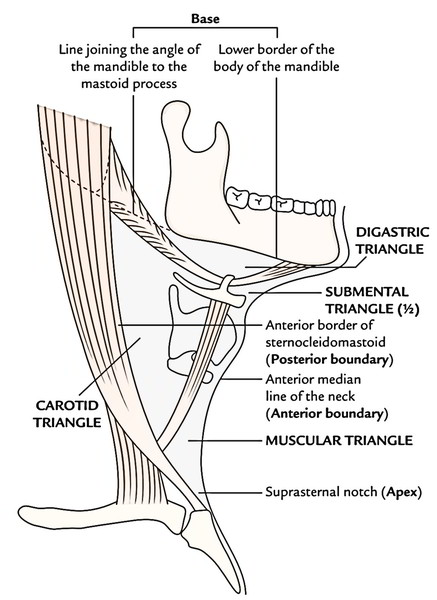
It is located on every side of the neck and is a large triangular space, with its apex pointed downwards and base pointed upwards and in front of sternocleidomastoid. 
Borders
- Anterior: Anterior median line of the neck.
- Posterior: Anterior border of the sternocleidomastoid muscle.
- Base: Lower border of the body of mandible and a lineextending from the angle of mandible to the mastoid process.
- Apex: Suprasternal notch in the meeting point betweenanterior border of sternocleidomastoid and anterior median line.
- Roof: Investing layer of deep cervical fascia.
The superficial fascia over the fascial roof includes platysma in the upper and anterior parts. The cervical branch of facial nerve and ascending and descending branches of transverse cervical cutaneous nerve traverse the plane between the platysma and the fascial roof of the triangle.
Subdivisions
For easy description, the anterior triangle hasbeen subdivided, by the digastric muscle and superior abdomen of omohyoid muscle, in the subsequent 3 1/2 triangles.
- Submental triangle (half only).
- Digastric (submandibular) triangle.
- Carotid triangle.
- Muscular triangle.
Before analyzing the subsections of anterior triangle, the awareness of the attachments of digastric and omohyoid muscles that are utilized for its subdivision is necessary. Digastric muscle: It has 2 abdomens- posterior and anterior. The triangular posterior abdomen from the digastric notch on the medial outermost layer of the base of the mastoid process originates and in the direction of the hyoid bone, runs downwards and forwards. The narrow anterior abdomen runs downwards and backwards and originates from digastric fossa on thebase of the mandible near symphysis menti, in the direction of the hyoid bone. Both midriffs are linked by an intermediatetendon, above the hyoid bone that is anchored to the hyoid bone by a fascial sling of investing layer of deep cervical fascia. Omohyoid muscle: It is composed of superior andinferior midriffs joined at an angle by an intermediate tendon. The inferior abdomen originates from upper border of scapula, nearthe suprascapular notch, it subsequently enters behind the sternocleidomastoid and ends there in an intermediatetendon. The superior abdomen starts at the intermediate tendon and enters nearly vertically upwards to be connected to thelower border of the hyoid bone. The intermediate tendon liesat the level of arch of cricoid cartilage and anchored to theclavicle by fascial sling originated from investing layer of deepcervical fascia.
Submental Triangle
It’s really a median triangle on the front of neck below the chin and above the hyoid bone. It therefore takes up the upper portions of both the anterior triangles. 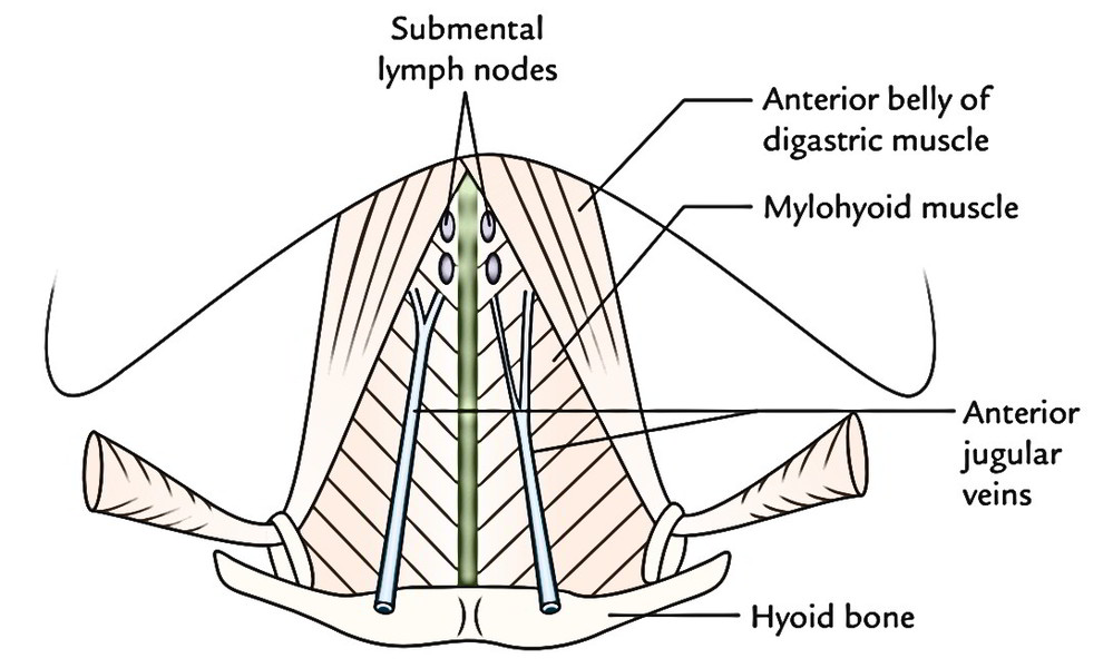
Borders
On every side: Anterior belly of digastric muscle.
- Base: Body of hyoid bone.
- Apex: Chin or symphysis menti.
Floor
It’s created by both mylohyoid muscles assembly at the median fibrous raphe stretching from symphysis menti to the hyoid bone.
Roof
Investing layer of deep cervical fascia.
Contents
- Submental lymph nodes (3 or 4 in number).
- Efferents from these nodes pass to the submandibular lymph nodes.
- Submental veins and commencement of anterior jugular veins.
- The contents of the triangle actually are located in the superficial fascia over the roof of the submental triangle.
Clinical Significance
Diseases of floor of mouth, tip of tongue and incisor teeth cause debilitating enlargement of submental lymph nodes because lymph from these structures is drained into these nodes.
Digastric (Submandibular) Triangle
It’s thus referred to as because it’s situated between both bellies of the digastric muscle and below the base of the mandible. 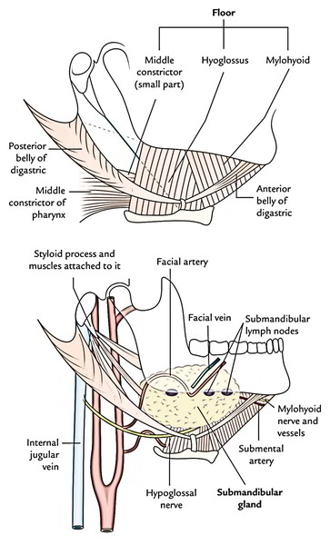
Borders
- Anteroinferiorly: Anterior belly of digastric muscle.
- Posteroinferiorly: Posterior belly of digastric muscle, supplemented by stylohyoid muscle.
- Base: It’s created by the base of the mandibleand imaginary line joining the angle of the mandible to the mastoid process.
- Apex: It’s created by the intermediate tendon ofthe digastric muscle, being bound down to hyoid bone by a fascial sling originated from investing layer of deep cervical fascia.
Floor
Roof
It’s created by investing layer of deep cervical fascia, which splits to enclose the submandibular salivary gland In the superficial fascia over the roof is located platysma, cervical branch of facial nerve and ascending branch of transverse cervical nerve.
Contents
The digastric triangle is subdivided into anterior and posterior parts by the stylomandibular ligament, which goes from the tip of the styloid process to the angle of the mandible. The posterior part of the triangle is constant above with the parotid region. Contents in the anterior part of the triangle:
- Submandibular salivary gland
- Submandibular lymph nodes.
- Hypoglossal nerve.
- Facial vein (is located superficial to the gland).
- Facial artery (is located deep to the gland).
- Submental artery.
- Mylohyoid nerve and boats.
Contents in the posterior part of the triangle:
- External carotid artery.
- Carotid sheath and its contents.
Structures passing between the external and internal carotid arteries.
Clinical Significance
Submandibular triangle is the common site of swellingsdue to: (a) engagement of submandibular lymph nodes as they drain lymph from prevalent regions like anterior two-third of tongue (with the exception of tip), floor of mouth, frontal, ethmoidal and maxillary air sinuses, etc. and (b) enlargement of the submandibular gland The efferents from submandibular lymph nodes largely pass to jugulo-omohyoid and partly to jugulodigastric nodes situated along the internal jugular vein.
Carotid Triangle
It’s so called because it includes all the 3 carotid arteries, viz. common carotid, internal carotid and external carotid. 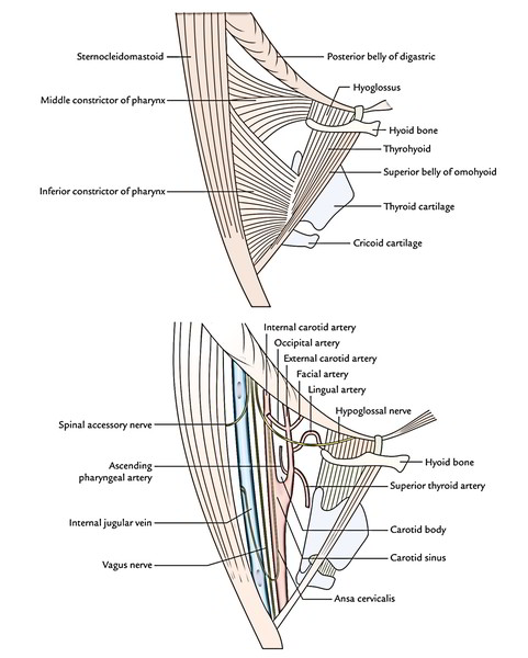
Bounds
- Superiorly: Posterior belly of digastric supplementedby stylohyoid.
- Anteroinferiorly: Superior belly of omohyoid.
- Posteriorly: Anterior border of sternocleidomastoid.
- Roof: It’s created by investing layer of deepcervical fascia. The superficial fascia over the roof includes platysma, cervical branchof facial nerve and transverse cervical nerve (a cutaneous branch of the cervical plexus).
- Floor: It’s created by the subsequent 4 muscles: Thyrohyoid, Hyoglossus, Middle constrictor and Inferior constrictor.
Contents
- Carotid arteries Common carotid artery, Internal carotid artery and External carotid artery and its first 5 branches.
- Carotid sinus and carotid body.
- Internal jugular vein.
- Last 3 cranial nerves Vagus nerve, Spinal accessory nerve and Hypoglossal nerve.
- Carotid sheath.
- Ansa cervicalis.
- Cervical part of the sympathetic chain.
- Deep cervical lymph nodes.
The major contents of carotid triangle are common carotid artery, internal carotid artery, external carotid artery, internal jugular vein and last 3 cranial nerves, all overlapped by sternocleidomastoid. The carotid triangle gives great view of all its contents only if sternocleidomastoid is retracted somewhat backwards.
Applicable Features of the Contents of the Carotid Triangle
Common Carotid Artery
It ascends from just behind the inferior angle of the carotid triangle in the carotid sheath under cover of anterior border of sternocleidomastoid and in front of lower 4 cervical transverse processes and in the level of the upper border of the thyroid cartilage breaks up into external and internal carotid arteries. The common carotid artery supplies no other branches in the neck.
Internal Carotid Artery
It runs straight upwards as the continuance of the common carotid artery The common carotid artery and its continuance, the internal carotid artery can be symbolized in living body by a straight line from sternoclavicular joint to the retromandibular fossa.
Clinical significance
The common carotid artery can be compressed against the notable anterior tubercle of transverse process of the 6th cervical vertebra termed carotid tubercle by pressing medially and posteriorly with the thumb. The carotid tubercle of the 6th cervical vertebra is all about 4 cm above the sternoclavicular joint.
External Carotid Artery
It ascends anteromedial to the internal carotid artery andgives the subsequent 5 branches in the triangle:
- Ascending pharyngeal artery (first branch).
- Superior thyroid artery.
- Lingual artery.
- Facial artery.
- Occipital artery.
- Ascending pharyngeal artery: It’s a slight artery that originates from the medial aspect of external carotid artery near its lower end. It ascends in the deeper plane on the side of the pharynx.
- Superior thyroid artery: It rises from the front and descends downwards and forwards to pass deep to the infrahyoid muscles to get to the upper part of the thyroid gland
- Lingual artery: It appears from the front aspect of external carotid artery, opposite the tip of greater cornu of hyoid bone. It then runs upwards and forwards creating a characteristic loop over the greater cornu of the hyoid and evaporate deep to the hyoglossus muscle. Before evaporating it supplies rise to suprahyoid artery, which runs superficial to hyoglossus above the hyoid.
- The loop of lingual artery above the greater cornu of hyoid bone allows free movements of hyoid bone without damaging the artery.
- Facial artery: It originates from the front aspect of external carotid artery just above the lingual artery and runs upwards on the superior constrictor deep to digastric and stylohyoid muscles.
- Occipital artery: It appears from the posterior aspect of external carotid artery at the lower border of posterior belly of digastric muscle and runs backwards superficial to internal carotid artery, internal jugular vein and last 3 cranial nerves along the lower border of posterior abdomen of digastrics.
Hypoglossal Nerve
It descends between the internal jugular vein and internal carotid artery. Afterward just above the level of greater cornu of hyoid bone, it hooks around the origin of the occipital artery, runs forwards, crossing in front of internal carotid artery, external carotid artery and loop of lingual artery to run on the hyoglossus muscle above the hyoid bone. As it crosses the internal carotid artery, it produces the superior root of ansa cervicalis (also termed descenholes hypoglossi), which descends on the anterior wall of the internal and common carotid arteries and becomes embedded in the anterior wall of the carotid sheath.
Deep Cervical Lymph Nodes
They be located on and along the internal jugular vein
Internal Jugular Vein
It’s partially concealed by the posterior edge of the sternocleidomastoid. It descends posterolateral to common and internal carotid arteries and gets the subsequent 3 veins in the region of carotid triangle:
- Lingual vein.
- Common facial vein.
- Superior thyroid vein.
The lingual vein is located just below and parallel to the hypoglossal nerve, crosses external and internal carotid arteries to join the internal jugular vein. The common facial vein after crossing the digastric triangle crosses the upper part of carotid triangle to drain into the internal jugular vein. The superior thyroid vein crosses the lower part of the triangle to finish into the internal jugular vein.
Vagus Nerve
It descends vertically downwards, first between the internal carotid artery and internal jugular vein and after that between common carotid artery and internal jugular vein. It supplies the subsequent 2 branches in the carotid triangle. Pharyngeal branch: It runs inferomedially between the external and internal carotid arteries to join the pharyngeal plexus on the superior constrictor of the pharynx. Superior laryngeal nerve: It runs on a deep plane, deep to both internal and external carotid arteries, where it breaks up into internal and external laryngeal nerves. The internal laryngeal nerve (sensory) enters forwards to evaporate deep to thyrohyoid muscle; there it pierces the thyrohyoid membrane to supplies the laryngeal mucosa. The external laryngeal nerve (motor) descends to supply the inferior constrictor and cricothyroid muscles after passing deep to the superior belly of the omohyoid.
Accessory Nerve
It runs downwards and backwards across the upper part of the triangle, superficial to the internal jugular vein to goes into the sternocleidomastoid muscle, which it furnishes.
Carotid Sheath
It’s a facial sheath which encloses internal jugular vein and internal and common carotid arteries. The vagus nerve is located between the vein and the artery on a deeper plane. The ansa cervicalis is embedded in its anterior wall on the other hand the cervical sympathetic chain is located just deep to its posterior wall on the prevertebral fascia.
Carotid Sinus
It’s a fusiform dilatation in the terminal end of common carotid artery or at the start of internal carotid artery. It’s rich innervation from glossopharyngeal, vagus and sympathetic nerves. The carotid sinus functions as a baroreceptor (pressure receptor) and modulates the blood pressure in the cerebral arteries. Clinical significance In an individual with carotid sinus hypersensitivity, pressure on carotid sinuses can cause enough slowing of heart rate, fall in blood pressure and cerebral ischemia that will bring about fainting (syncope). People with hypersensitive carotid sinuses regularly had unexpected episodes of syncope on rotation of head particularly when wearing a top with tight collar or a tie with tight knot, ailment named carotid sinus syndrome. Symptoms can be alleviated by periarterial neurectomy.
Carotid Body
It’s a small oval structure situated behind the bifurcation of the common carotid artery. It’s reddish brown and gets abundant Nerve Supply from glossopharyngeal, vagus and sympathetic nerves. It acts as a chemoreceptor and reacts to the changes in the oxygen and carbon dioxide content of the blood. The excess of CO2 and reduced O2 tension in blood sparks it causing increase in blood pressure and heart rate.
Ansa Cervicalis
The ansa cervicalis (ansa hypoglossi) is a U-shaped nerve loop within the region of the carotid triangle embedded in the anterior wall of the carotid sheath. It’s originated from ventral rami of C1, C2 and C3 spinal nerves. It supplies all the infrahyoid muscles with the exception of thyrohyoid that is supplied by nerve to thyrohyoid (C1) from hypoglossal nerve. Roots: Ansa cervicalis has the following 2 roots:
- Superior root (descenholes hypoglossi) is composed by the descending branch of the hypoglossal nerve taking C1 spinal nerve fibres. It descends downwards over internal and common carotid arteries.
- Inferior root (descenholes cervicalis) is originated from C2 and C3 spinal nerves. As this root descends, it first winds round the internal jugular vein and after that continues anteroinferiorly to join the superior root in front of the common carotid artery in the level of cricoid cartilage.
Distribution:
- Superior root supplies branch to superior belly of omohyoid.
- Dependent loop supplies branches to sternohyoid, sternothyroid and inferior belly of the omohyoid.
Muscular Triangle
Borders
Anterior: Anterior median line of the neck, going from hyoid bone to the suprasternal notch. Anterosuperior: Superior belly of the omohyoid. Posteroinferior: Anterior border of sternocleidomastoid.
Floor
It’s created by sternothyroid, sternohyoid and thyrohyoid muscles.
Roof
It’s created by investing layer of deep cervical fascia. The superficial fascia over the roof includes anterior jugular vein and related lymph nodes.
Contents
- Infrahyoid muscles, viz. sternothyroid, sternohyoid and thyrohyoid.
- The significant deeper structures in the region of muscular triangle are thyroid gland, trachea and esophagus. They’re studied individually.
- The infrahyoid area includes strap (ribbon like) muscles of the neck. Underneath these muscles is located all important structures. From superficial to deep, these are:
- Thyroid and parathyroid glands.
- Larynx and trachea.
- Hypopharynx (laryngopharynx) and esophagus.
Key Points: Supra and infrahyoid regions: All these really are the regions on the front of neck above and below the hyoid bone, respectively. Suprahyoid area corresponds to submental triangle. Infrahyoid area is bounded superolaterally by the superior abdomens of the omohyoid muscles and inferolaterally by the sternocleidomastoid muscles. It consists of muscular triangles of both anterior triangles.


 (86 votes, average: 3.50 out of 5)