The Head is the globe shaped ,topmost portion of the body, that consists of the skull, brain and sensory organs that includes , eyes that allows to have a vision, ears for sensing sounds and maintaining equilibrium (balance), nose that identifies and senses various types of smell, and tongue that identifies and senses tastes. The head also provides openings for respiratory and digestive systems, that also acts as a starting point. The head can be divided into two portions; the cranium and the face.
The cranium (a.k.a the skull, the braincase etc) consists of brain. The openings for eyes , nose, and mouth oral cavity is located in face.
A little description of comparative anatomy makes the differentiation between the size of cranium and face simpler to understand.
The sense of smell is just one of the earliest sensitivities. The pronograde canines (example, dog) are directed primarily by smell for hunting food and sex. The other senses, like touch, hearing, and eyesight play an accessory function. For that reason, they have well-developed snout, and, their face lies in front of the cranium.
The arboreal way of life of apes and monkeys favored the higher development of visual, acoustic, tactile, kinesthetic, and motor functions with advancement in their own brains. In such creatures, utility of the nose was lost and sense of smell became an accessory perception. Therefore in orthograde monkeys, it resulted in the loss of the projecting snout, and there face is situated below and in front of the cranium.
The supremacy of guy in animal kingdom is because of his large well-developed brain, which gives him the infinite power of thinking, reasoning, and judgement. To adapt large brain, the size of cranium has also grown proportionately. Thus, in plantigrade guy the forehead is notable and the face lies below the anterior part of the cranium.
It’s significant to notice that size of jaws is inversely proportional to the size of cranium. Hence the pronograde canine has bigger jaws; an orthograde monkey has smaller jaws on the other hand plantigrade guy has smallest jaws. The reduction in the size of jaws happened as a result of shift in eating habits of these creatures. The jaws are smallest in guy because he prefers to eat soft cooked food. The size of jaws is bigger in canines since they use it for holding, breaking, biting, snapping, and chewing the food. With receding jaws, the mouth is proportionately reduced in size.
In man, eyes are positioned in more frontal plane to allow stereoscopic vision. To allow freedom of mobility to the tongue for a well-articulated language in guy, the alveolar arches are extended and the chin is pushed forward, making the mouth cavity more roomy. The notable chin is a characteristic attribute of human beings. The identifying external nose with notable dorsum, tip, and alae is characteristic of a guy, in spite of the fact that it doesn’t have anything related to the sense of smell. Likely it functions to shield the eyes from injuries. The brow ridges are noticeably reduced in man as compared to other primates as a result of their notable forehead.

Living Anatomy
The living anatomy addresses the evaluation of surface features by visualization (review) and palpation of the living people to get info regarding the deeper structures. It’s of tremendous value in clinical examination of the patients. The study of living anatomy (also termed living or surface anatomy) of head and neck starts with the section of the surface into regions and examining surface landmarks in every region. The people are counselled to practice finding these landmarks in every region on themselves or on their co-workers to acquire the art of evaluation.
Regions of The Head
The head is split into these regions: frontal, parietal, occipital, temporal, auricular, parotid, orbital, nasal, zygomatic, buccal, oral, and mental.
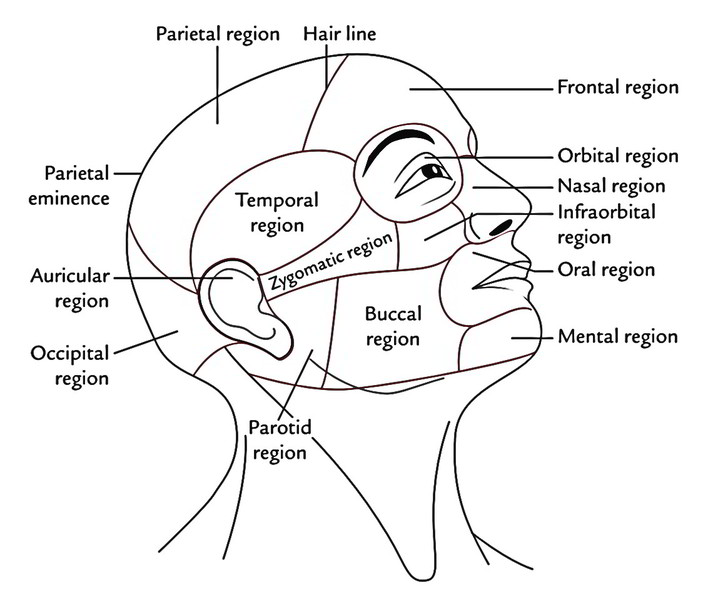
Frontal Region (Brow)
The frontal region of the head is an area above the eyes and below the hair line. Eyebrows are the raised arches of skin with short, thick hairs above the supraorbital margins. Just deep to eyebrow is the curved bony ridge or superciliary arch. It’s more notable in adult males. The smooth non-hairy raised area between the eyebrows is named glabella, which tends to be flat in kids and adult females, and creates a rounded bulge in adult males. Indian wed Hindu females employ bindi at this site to improve their attractiveness. It’s essential to notice the pineal gland is located about 7 cm behind the glabella. The bulge of forehead, the frontal eminence is clear on either side above the eyebrow. The frontal bulge is normally more marked in youngsters and adult females.
Parietal Region
It’s an area restricted anteriorly by hair line and posteriorly by a coronal plane behind the parietal eminences and on each side by the temporal line. The parietal eminence can be felt on each side in this region about 2 inches above the auricle. The parietal bulges are clear on or just in front of the interauricular line.
Occipital Region
The occipital region is an area of cranium behind the parietal eminences, and above the external occipital protuberance and superior nuchal lines.
The most notable point in the occipital region is referred to as opisthocranion or occiput. The external occipital protuberance can be felt in the median line just above the nuchal furrow. The superior nuchal line, 1 on either side of external occipital protuberance, runs laterally with its convexity facing upwards.
The soft tissue covering frontal, parietal, and occipital regions creates the scalp.
Clinical Significance
The large area of scalp over the vault of skull is thickly covered by terminal hair. Because of presence of hair, many lesions in this area stay undetected by both clinicians and patients. Therefore, this area ought to be cautiously analyzed by the clinicians.
Temporal Region (Temple)
The temporal region is the area on the side of the skull between the temporal line and zygomatic arch.
It’s the site of connection of temporalis muscle, which can be palpated when the teeth are clenched repeatedly. Attempt on yourself. Soft tissue in the temporal region consists of skin, subcutaneous tissue, temporal fascia, and temporalis muscle. In the anterior part of temporal region, deep to soft tissues is a small area where 4 bones meet the pterion. This region is medically significant because it’s the site of entry to cranial cavity in craniotomy to eliminate the extraduralhematoma.
Auricular Region
The auricular region consists of fleshy oval flap of the ear (auricle) and external acoustic meatus.
The auricle collects the sound waves. The external auditory meatus is a tube via which sound waves are carried to the middle ear inside the skull. Detect the following surface features of the auricle.
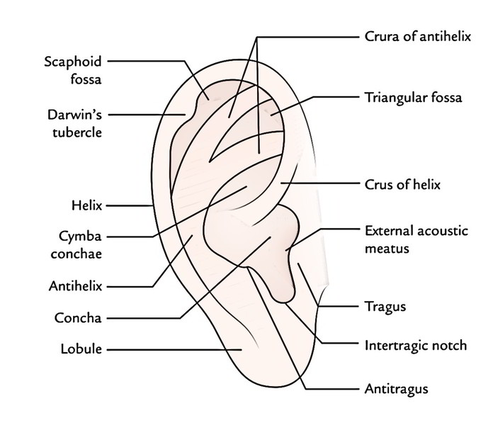
The superior and posterior free margins of the auricle creating a type of rim are referred to as helix, which finishes inferiorly at the fleshy protuberance of the ear named ear lobule.
The upper end of the helix is generally at the level of the eyebrows and the glabella.
The lobule is roughly at the level of the apex of the nose.
The portion of the auricle anterior to the external auditory meatus is a small nodular flap of tissue termed tragus. It projects posteriorly, partly covering and shielding the external auditory meatus. The condyle of mandible can be palpated by placing the tip of finger just in front of tragus and after that opening and shutting the mouth.
Another flap of tissue opposite the tragus is the antitragus. Between the tragus and antitragus is a deep notch named awful notch.
A semicircular ridge anterior to the helix is referred to as antihelix.
The upper end of antihelix splits into 2 crura enclosing a triangular depression named triangular fossa. The depressed hollow of the auricle is termed concha.
The upper end of the helix which goes backwards to some extent into concha is termed crux of helix.
Clinical Significance
The external auditory meatus and tragus are significant landmarks to work with when shooting extraoral radiographs and administering local anesthesia on a patient. The pulsations of superficial temporal artery can be felt by placing the fingertip just in front and above the tragus on the root of zygoma.
Parotid Region
As the particular name suggests, it’s present around the ear (para = around; otic = ear). It’s limited in front by anterior border of masseter, behind by mastoid process and below by line stretching from angle of mandible to the tip of mastoid process. This region is inhabited by parotid gland. The mastoid process is located behind the lower part of the ear. Its anterior border, tip and posterior border can be easily felt.
The masseter overlies the ramus of the mandible. It can be felt when the teeth are clenched.
Clinical Significance
The parotid gland is frequently enlarged following infection by mumps virus. This creates a painful swelling in the parotid region elevating the ear lobule. The parotid gland is also the site of slow growing painless tumor termed combined parotid tumor.
Orbital (Ocular) Region
The ocular region consists of the eyeball and related structures. Majority of the surface features of the ocular region protect the eye. Eyebrow is a ridge of hair along the superciliary arch above the orbit, which protects the eyes against sun and mechanical setback. Both movable eyelids reflexly close to shield eyes from foreign particles and bright sun. The eyelashes are a row of hair in the margins of eyelids. The eye-lashes prevent airborne things from contacting the eyeball. Behind the lateral part of the upper eyelid and inside the orbit is the lacrimal gland, which generates lacrimal fluid or tears. The tears wash away chemical and foreign particles and lubricate the front of the eye to forbid the face of the eyeball, especially the all-important cornea from drying.
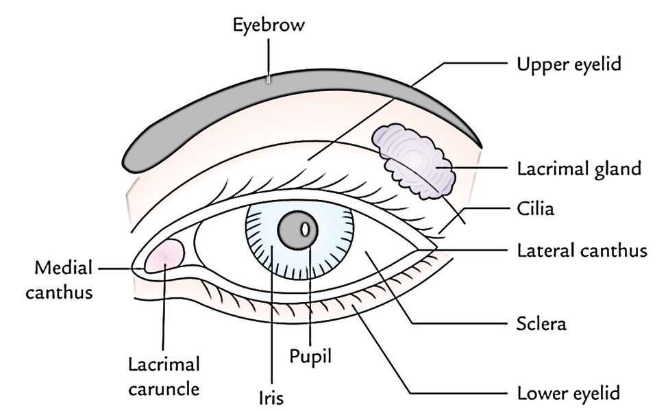
The conjunctiva is a fine thin mucous membrane which lines the inner surface of the eyelids and the front of the eyeball. It assists in reducing friction during blinking.
The sclera, the ‘white’ of the eye is viewed on each side of cornea.
The cornea is the circular clear anterior portion of the eyeball.
The outer corner where the upper and lower eyelids meet is named lateral (outer) canthus. The inner corner where the 2 eyelids meet is referred to as medial canthus. A fleshy pinkish elevation in the medial angle of the eye is referred to as lacrimal caruncle.
Palpate these landmarks in this region in yourself:
- Supraorbital pass on the maximal point of supraorbital margin about 2.5 cm from the midline.
- Frontozygomatic suture, that is marked by a slight atypical depression on the lateral orbital margin.
Clinical Significance
The status of the eyes deeply changes the facial look. Lesions affecting the eye and its related structures are tremendous. A few easily identifiable and surgically important circumstances are as follows:
- Arcus senilis, a white rim around the outer edge of the iris, is often observed in aged individuals. It takes place because of sclerosis and deposit of cholesterol in the boundary of the cornea.
- Xanthelasma are fatty plaques in the skin of the eyelids. They look like masses of yellowish opaque fat. If multiple and growing, they signify inherent abnormality of cholesterol metabolism, diabetes, or arterial disease.
- Exophthalmos is a forward protrusion of the eyeball from its normal position in the orbit. The commonest cause of both bilateral and unilateral exophthalmos is thyrotoxicosis (hyperthyroidism).
- Ectropion is the eversion of the lower eyelid.
Nasal Region
The key attribute of nasal region is the external nose. It’s a pyramidal projection in the middle third of the face with its root up and base downwards. The root of the nose can be found between the eyes inferior to glabella. The solid narrow bony portion below the nasion is the bridge of the nose. The nose below this level has pliable cartilaginous framework that keeps the openings of the nose. The tip of the nose is named apex. It’s flexible when palpated because it’s created from cartilage. Inferolateral to the apex on each side is a nostril (or nare). The nostrils are divided from every other by a midline nasal septum. The nares are bounded laterally by wing like alae of the nose. The alae of nose create the flared outer margin of every nostril.
The identifying external nose with exuberant development of cartilages creating notable dorsum, tip, and alae is a characteristic attribute of human beings.
A well-marked depression in the root of the nose is referred to as nasion.
Clinical Significance
Saddle nose: A nose whose bridge is depressed and widened.
Rhinophyma: The nasal skin covering the alar cartilages is thick and adherent, and includes many sebaceous glands. The hypertrophy and adenomatous changes of these glands provides rise to a lobulated tumor termed rhinophyma.
Infraorbital Region
The infraorbital region of head is situated below the orbital region and corresponds to the upper part of the anterior surface of the maxilla. The infraorbital foramen lies in this region about 1 cm below the infraorbital margin consistent together with the supraorbital notch or foramen. The knowledge of its location is essential for providing infraorbital nerve block.
Zygomatic Region
The zygomatic region overlies the zygomatic (cheek) bone and zygomatic arch.
The zygomatic arch stretches from just inferior to lateral margin of the eye in the direction of the upper portion of the auricle. Inferior to the zygomatic arch and just anterior to the tragus of the ear is the temporomandibular joint. The zygomatic arch is bony bridge that covers the interval between the ear and the eye. The zygomatic bone creates the bony prominence of the cheek below and lateral to the orbit.
The movements of the temporomandibular joint can be felt by opening and shutting the mouth or moving the lower jaw from side to side. 1 way to sense the movements of head of mandible will be to gently put a finger into the outer portion of the external auditory meatus.
Buccal Region
The buccal region of face is a broad area of the face between the nose, mouth, and parotid region. It overlies the buccinator muscle. It’s made of soft tissues of the cheek.
The pulsations of facial artery can be felt about 1.25 cm lateral to the angle of the mouth.
Oral Region
The structures of the oral region contain fleshy upper and lower lips, and the structures of oral cavity that can be discovered when the mouth is broadly open.
The lips are mainly composed of muscles covered exter-nally by skin and internally by mucous membrane. Every lip has a pinkish zone named vermillion zone. The lips are outside-lined from the surrounding skin by a transition zone named vermillion border. The small triangular median depression in the upper lip is termed philtrum. The apex of philtrum is in the direction of the nasal septum and the base downwards where it ends in a thicker area named tubercle of the upper lip.
Clinical Significance
The corners of mouth where upper and lower lips meet are referred to as labial commissure. The groove running upward between the labial commissure and the alae of nose is known as nasolabial sulcus. The lower lip is divided from the chin by a horizontal groove named labiomental groove.
The colour of the lips and the mucus membrane of the oral cavity are medically significant; lips may seem pale in patients with acute anemia or bluish in individuals afflicted by dearth of oxygenation of blood (cyanosis). A lemon yellow tint of lips may signify jaundice.
The lips are a standard site for carcinoma, mainly impacting people above 60 years old. Carcinoma of the lip generally takes place in lower lip (93%) as compared to the upper lip (5%).
The bone underlying the upper lip is the alveolar process of the maxilla, on the other hand the bone underlying the lower lip is the alveolar process of the mandible. The alveolar processes include teeth and are referred to as maxillary and mandibular teeth.
Mental Region
The mental region is an area of face below the lower lip and is qualified by the presence of mental protuberance or mentum, a privileged attribute of human beings.
- The part of oral cavity within the alveolar arches is named oral cavity appropriate. It includes a mobile muscular organ, the tongue.
- The oral cavity is lined by a mucus membrane or mucosa. The inner aspects of the lips are lined by pink and thick labial mucosa. The labial mucosa is constant with the equally pink and thick buccal mucosa that lines the inner cheek.
- The space between cheek/lip and gum is known as vestibule.
- On the inner aspect of buccal mucosa opposite the upper second molar tooth is a small elevation referred to as parotid papilla on which opens the parotid duct.
- The gingiva is a part of oral mucosa that covers the alveolar processes of the jaws.
- The roof of oral cavity which presents 2 portions: (a) a solid anterior portion is termed hard palate and a flexible posterior portion is named soft palate. A cone shaped projection hanging down from the middle of the posterior free margin is named uvula of the palate, that is constant with palatopharyngeal arch on every side.
- A dense pad of soft tissue behind the final molar tooth is termed retromolar pad.
- The floor of mouth lies inferior to the ventral surface of the tongue.
The oral cavity gives entry into the throat or thepharynx.
One can easily analyze these features in theoropharynx:
A curved, leaf-like flap of cartilage lies behind the base of tongue and in front of oropharynx. It’s epiglottis, the cartilage of the larynx.
Mass of lymphoid tissue projecting on either side into the lateral wall of the oropharynx is termed palatine tonsil. The palatine tonsils are by and large named tonsils by the patients. The tonsil is located in triangular fossa named tonsillar fossa found between the palatoglossal and palatopharyngeal arches. Notice the tonsils be located opposite the angle of mandible between the rear of tongue and soft palate.
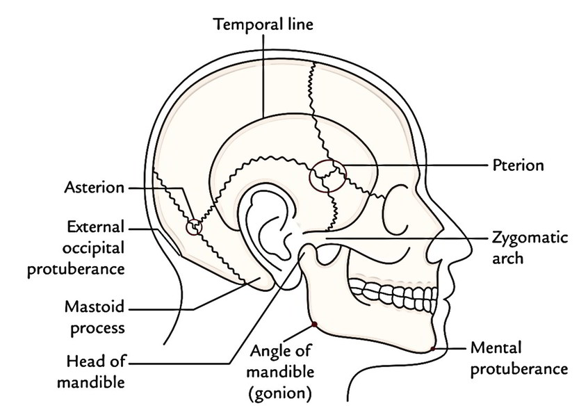
Bony Landmarks In The Region of Head
| Landmark | Location |
|---|---|
| Mental protuberance/ mentum | Protuberance of the chin |
| Nasion | Depression at the root of nose at the junction of frontonasal and internasal sutures |
| Glabella | Smooth non-hairy area between the eyebrows above nasion |
| Vertex | Highest point on the top of head in the midline |
| External occipital protuberance | Knob-like bony projection at the upper end of nuchal furrow |
| Inion | Apex of external occipital protuberance |
| Gonion | Angle of mandible |
| Head of mandible | In front of lower part of the tragus |
| Preauricular point | In front of upper part of the tragus |
| Mastoid process | Behind the lower part of the auricle |
| Pterion | 4 cm above the midpoint of zygomatic arch/3.5 cm behind and 1.5 cm above the frontozygomatic suture |
| Asterion | Depression—2.5 cm behind the upper part of the root of ear |
| Supraorbital notch/foramen | On the supraorbital margin 2.5 cm from midline |
| Infraorbital foramen | 1 cm below infraorbital margin and 1.25 cm lateral to the side of nose |
| Mental foramen | 2.5 cm lateral to symphysis menti and 1.25 cm above the lower border of mandible |
| Frontal prominence | Area of maximum convexity on either side of forehead where top, front and side of head meet |
| Parietal prominence | Area of maximum convexity on either side in the parietal region where back, top and side of head meet (Area of maximum transverse diameter of the skull) |

 (59 votes, average: 4.66 out of 5)