There are more than 600 muscles in the body, but only a few of the major muscles are considered here.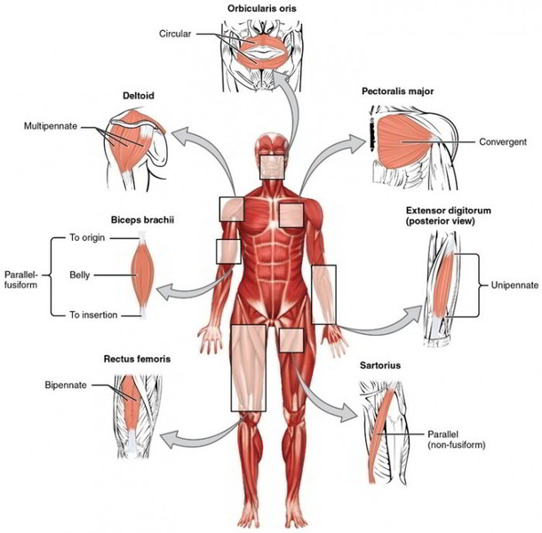
Muscles of Facial Expression And Mastication
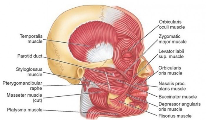
Muscles of the face and scalp produce the facial expressions that help communicate feelings, such as anger, sadness, happiness, fear, disgust, pain, and surprise. Most have origins on skull bones and insertions on the dermis of the skin.
| Muscle | Origin | Insertion | Action |
| Buccinator | Lateral surfaces of maxilla and mandible | Orbicularis oris | Compresses cheeks inward |
| Epicranius | This muscle consists of two parts: the frontal belly and the occipital belly. They are joined by the epicranial aponeurosis, which covers the top of the skull. | ||
| Frontal belly | Epicranial aponeurosis | Skin and muscles superior to the eyes | Elevates eyebrows and wrinkles forehead |
| Occipital belly | Base of occipital bone | Epicranial aponeurosis | Pulls scalp posteriorly |
| Orbicularis oculi | Frontal bone and maxillae | Skin around eye | Closes eye |
| Orbicularis oris | Muscles around mouth | Skin around lips | Closes and puckers lips; shapes lips during speech |
| Platysma (plah-tiz’-mah) | Fascia of superior chest | Mandible and muscles around mouth | Draws angle of mouth inferiorly |
| Zygomaticus | Zygomatic bone | Orbicularis oris at angle of the mouth | Elevates corners of mouth (smiling) |
The epicranius is an unusual muscle. It has a large epicranial aponeurosis that covers the top of the skull and two contractile portions: the frontal belly over the frontal bone and the occipital belly over the occipital bone.
Two major pairs of muscles elevate the mandible in the process of mastication (chewing): the masseter and the temporalis.
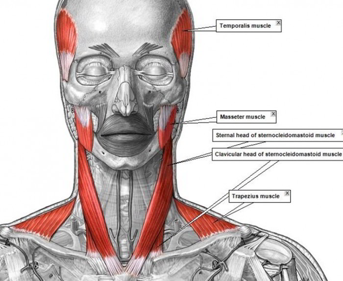
Muscles That Move The Head
Several pairs of neck muscles are responsible for flexing, extending, and rotating the head. Table below lists two of the major muscles that perform this function: the sternocleidomastoid and the splenius capitis. The trapezius can also extend the head, although this is not its major function.
| Muscle | Origin | Insertion | Action |
| Sternocleidomastoid | Clavicle and sternum | Mastoid process of temporal bone | Contraction of both muscles flexes head toward chest; contraction of one muscle turns head away from contracting muscle |
| Splenius capitis | Inferior cervical and | Mastoid process | Contraction of both muscles extends head; |
| superior thoracic vertebrae | of temporal bone | contraction of one muscle turns head toward same side as contracting muscle |
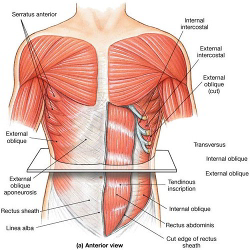
Muscle of The Abdominal Wall
The abdominal muscles are paired muscles that provide support for the anterior and lateral portions of the abdominal and pelvic regions, including support for the internal organs. The muscles are named for the direction of their muscle fibers: rectus abdominis, external oblique, internal oblique, and transversus abdominis. They are arranged in overlapping layers and are attached by larger aponeuroses that merge at the anterior midline to form the linea alba, or white line.
| Muscle | Origin | Insertion | Action |
| Rectus abdominis | Pubic symphysis and pubis | Xiphoid process of sternum and costal cartilages of ribs 5 to 7 | Tightens abdominal wall; flexes the vertebral column |
| External oblique | Anterior surface of inferior eight ribs | Iliac crest and linea alba | Tightens abdominal wall; rotation and lateral flexion of the vertebral column |
| Internal oblique | Iliac crest and inguinal ligament | Cartilage of inferior four ribs, pubis, and linea alba | Same as above |
| Transversus abdominis | Iliac crest, cartilages of inferior six ribs, processes of lumbar vertebrae | Pubis and linea alba | Tightens abdominal wall |
Muscles of Breathing
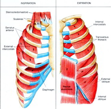
Movement of the ribs occurs during breathing and is brought about by the contraction of two sets of muscles that are located between the ribs. The external intercostals elevate and protract the ribs during inspiration, and the internal intercostals depress and retract the ribs during expiration. The primary breathing muscle is the diaphragm, a thin sheet of muscle that separates the thoracic and abdominal cavities.
| Muscle | Origin | Insertion | Action |
| Diaphragm | Lumbar vertebrae, costal | Central tendon | Forms floor of thoracic cavity; |
| cartilages of inferior ribs. | located at midpoint | depresses during contraction. | |
| xiphoid process | of muscle | causing inspiration | |
| External intercostals | Inferior border of | Superior border | Elevates and protracts ribs |
| rib above | of rib below | during inspiration | |
| Internal intercostals | Superior border of rib | Inferior border | Depresses and retracts ribs |
| below | of rib above | during expiration |
Muscles That Move The Pectoral Girdle
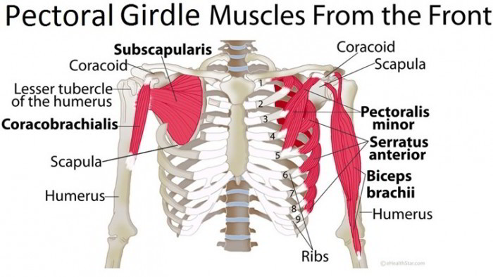
Pectoral girdle muscles originate on bones of the axial skeleton and insert on the scapula or clavicle. Because the scapula is supported mainly by muscles, it can be moved more freely than the clavicle. The trapezius is a superficial trapezoid-shaped muscle that covers much of the superior back. The rhomboid major and minor and the ievator scapulae lie deep to the trapezius. Each serratus anterior is located on the lateral surface of the superior ribs near the axillary region. The pectoralis minor lies deep to the pectoralis major. It protracts and depresses the scapula.
| Muscle | Origin | Insertion | Action |
| Trapezius | Occipital bone; cervical and thoracic vertebrae | Clavicle; spine and acromion of scapula | Elevates clavicle; adducts and elevates scapula; extends head |
| Rhomboid major and minor | Superior thoracic vertebrae | Medial border of scapula | Adducts and elevates scapula |
| Levator scapulae | Cervical vertebrae | Superior medial margin of scapula | Elevates scapula |
| Serratus anterior | Superior eight to nine ribs | Medial border of scapula | Depresses, protracts, and rotates scapula |
| Pectoralis minor | Anterior surface of superior ribs | Coracoid process of scapula | Depresses and protracts scapula |
Muscles That Move The Arm And Forearm
Movement of the humerus is enabled by the muscles that originate on the pectoral girdle, ribs, or vertebrae and insert on the humerus. The arrangement of these muscles and the ball-and-socket joint between the humerus and scapula enable great freedom of movement for the arm. The pectoralis major is the large superficial muscle of the chest. The deltoid is the thick muscle that caps theshoulder joint. The supraspinatus, infraspinatus, and teres minor cover the posterior surface of the scapula.
The anterior surface of each scapula is covered by the subscapularis. These four muscles and their tendons surround the head of the humerus at the shoulder joint, making up the rotator cuff The muscles and tendons of the rotator cuff are the only structures stabilizing the shoulder joint; thus the joint is fairly unstable compared to other joints. However, this relative lack of stability is what allows the shoulder’s mobility. The fatissimus dorsi is a broad, sheetlike muscle that covers the inferior back. The teres major assists the latissimus dorsi and is located just superior to it.
Muscles moving the forearm originate on either the humerus or the scapula and insert on either the radius or the ulna. Three flexors occur on the anterior surface of the arm: the biceps brachii, brachialis, and brachioradialis. One extensor, the triceps brachii, is located on the posterior surface of the arm.
Arms
| Muscle | Origin | Insertion | Action |
| Pectoralis major | Clavicle, sternum, and cartilages of superior ribs | Greater tubercle of humerus | Adducts, flexes, and medially rotates arm |
| Deltoid | Clavicle and spine, and acromion of scapula | Deltoid tuberosity of humerus | Abducts, flexes, and extends arm |
| Latissimus dorsi | Inferior thoracic and lumbar vertebrae; sacrum; inferior ribs; iliac crest | Intertubercular sulcus of humerus | Adducts, extends, and medially rotates arm |
| Teres major | Inferior angle of scapula | Distal to lesser tubercle of humerus | Same as above |
| Rotator cuff muscles | These four muscles stabilize the shoulder joint | ||
| Supraspinatus | Superior to spine of scapula | Greater tubercle of humerus | Abducts arm |
| Infraspinatus | Inferior to spine of scapula | Greater tubercle of humerus | Laterally rotates arm |
| Teres minor | Lateral border of scapula | Greater tubercle of humerus | Laterally rotates arm |
| Subscapularis | Anterior surface of scapula | Lesser tubercle of humerus | Medially rotates arm |
Forearms
| Muscle | Origin | Insertion | Action |
| Biceps brachii | Coracoid process and tubercle superior to glenoid cavity of scapula | Radial tuberosity of radius | Flexes forearm and supination, also flexes arm |
| Brachialis | Distal, anterior surface of humerus | Coronoid process of ulna | Flexes forearm |
| Brachioradialis | Lateral surface of distal end of humerus | Lateral surface of radius superior to styloid process | Flexes forearm |
| Triceps brachii | Lateral and medial surfaces of humerus and tubercle inferior to glenoid cavity of scapula | Olecranon of ulna | Extends forearm, also extends arm |
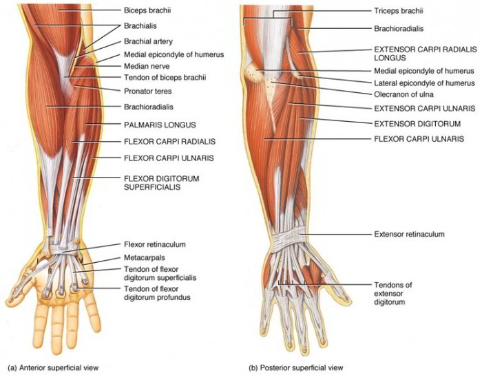
Muscles That Move The Wrist And Fingers
Many muscles that produce the various movements of the wrist and fingers are located in the forearm. Only a few of the larger superficial muscles are considered here. They originate from the distal end of the humerus and insert on carpal bones, metacarpals, or phalanges. Flexors on the anterior surface include the flexor carpi radialis, flexor carpi ulnaris, and palmaris longus. Extensors on the posterior surface include the extensor carpi radialis longus, extensor carpi ulnaris, and extensor digitorum (table 7.11; and figures 7.18 and 7.19). Note that the tendons of these muscles are held in position by a circular ligament at the wrist. Muscles That Move the Wrist and Fingers
| Muscle | Origin | Insertion | Action |
| Flexor carpi radialis | Medial epicondyle of humerus | Metacarpals II and III | Flexes and abducts wrist |
| Flexor carpi ulnaris | Medial epicondyle of humerus and olecranon of ulna | Carpal bones and metacarpal V | Flexes and adducts wrist |
| Palmaris longus | Medial epicondyle of humerus | Fascia of palm | Flexes wrist |
| Extensor carpi radialis longus | Lateral epicondyle of humerus | Metacarpal II | Extends and abducts wrist |
| Extensor carpi ulnaris | Lateral epicondyle of humerus | Metacarpal V | Extends and adducts wrist |
| Extensor digitorum | Lateral epicondyle of humerus | Posterior surfaces of phalanges II—V | Extends fingers |
Muscles That Move The Thigh And Leg
Muscles moving the thigh span the hip joint. They insert on the femur, and most originate on the pelvic girdle. The iliacus and psoas major are located anteriorly, the gluteus maximus is located posteriorly and forms the buttocks, the gluteus medius is located deep to the gluteus maximus posteriorly and extends laterally, and the tensor fasciae latae is located laterally. The adductor longus and adductor magnus are both located medially.
The leg is moved by muscles located in the thigh. They span the knee joint and originate on the pelvic girdle or femur and insert on the tibia or fibula. The quadriceps femoris is composed of four muscles that have a common tendon that inserts on the patella. However, this tendon continues as the patellar ligament, which attaches to the tibial tuberosity-the functional insertion for these muscles. The biceps femoris, semitendinosus, and semimembranosus on the posterior surface of the thigh are often collectively called the hamstrings. The medially
located gracilis has two insertions that give it dual actions. The long, straplike sartorius extends diagonally across the anterior surface of the thigh and spans both the hip and knee joints. Its contraction enables the legs to cross.
Thigh
| Muscle | Origin | Insertion | Action |
| Iliacus | Fossa of ilium | Lesser trochanter of femur | Flexes thigh |
| Psoas major | Lumbar vertebrae | Lesser trochanter of femur | Flexes thigh |
| Gluteus maximus | Posterior surfaces of ilium, sacrum, and coccyx | Posterior surface of femur and iliotibial tract | Extends and laterally rotates thigh |
| Gluteus medius | Lateral surface of ilium | Greater trochanter of femur | Abducts and medially rotates thigh |
| Tensor fasciae latae | Anterior iliac crest | Iliotibial tract | Flexes and abducts thigh |
| Adductor longus | Pubis near pubic symphysis | Posterior surface of femur | Adducts, flexes, and laterally rotates thigh |
| Adductor magnus | Inferior portion of ischium and pubis | Same as above | Same as above |
Leg
| Muscle | Origin | Insertion | Action |
| Quadriceps femoris | Four muscles of the anterior thigh that extend the leg. | ||
| Rectus femoris | Anterior inferior iliac spine and | Patella; tendon continues | Extends leg and flexes thigh |
| superior margin of acetabulum | as patellar ligament, which attaches to tibial tuberosity | ||
| Vastus lateralis | Greater trochanter and posterior surface of femur | Same as above | Extends leg |
| Vastus medialis | Medial and posterior surfaces of femur | Same as above | Extends leg |
| Vastus intermedius | Anterior and lateral surfaces of femur | Same as above | Extends leg |
| Hamstrings | Three distinct muscles of the posterior thigh that flex leg and extend thigh. | ||
| Biceps femoris | Ischial tuberosity and posterior | Head of fibula and lateral | Flexes and laterally rotates |
| surface of femur | condyle of tibia | leg; extends thigh | |
| Semitendinosus (sem-e-ten-di-no’-sus) | Ischial tuberosity | Medial surface of tibia | Flexes and medially rotates leg; extends thigh |
| Semimembranosus (sem-e-mem-brah-no’-sus) | Ischial tuberosity | Medial condyle of tibia | Flexes and medially rotates leg; extends thigh |
| Gracilis (gras’-il-is) | Pubis near pubic symphysis | Medial surface of tibia | Adducts thigh; flexes leg and locks knee |
| Sartorius (sar-to’r-e-us) | Anterior superior iliac spine | Medial surface of tibia | Flexes thigh and leg; abducts and laterally rotates thigh |
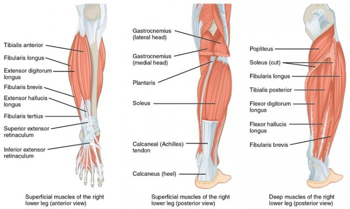
Muscles That Move The Foot And Toes
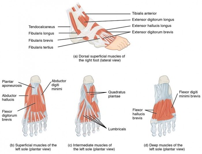 Many muscles are involved in the movement of the foot and toes. They are located in the leg and originate on the femur, tibia, or fibula and insert on the tarsal bones, metatarsals, or phalanges. The posterior leg muscles include the gastrocnemius and soleus, which insert through a common tendon, the calcaneal (Achilles) tendon, which attaches to the calcaneus. The tibialis anterior is anteriorly located, and the extensor digitorum longus lies lateral to it. Note that although the extensor digitorum extends the toes, as its name implies, it also dorsiflexes the foot. The fibularis longus is located on the lateral surface of the leg.
| Muscle | Origin | Insertion | Action |
| Gastrocnemius | Medial and lateral condyles of femur | Calcaneus by the calcaneal tendon | Plantar flexes foot and flexes leg |
| Soleus (so’l-e-us) | Posterior surface of tibia and fibula | Calcaneus by the calcaneal tendon | Plantar flexes foot |
| Fibularis longus | Lateral condyle of tibia and | Metatarsal I and tarsal | Plantar flexes and everts foot; |
| head and body of fibula | bones | supports arch | |
| Tibialis anterior | Lateral condyle and surface of tibia | Metatarsal I and tarsal bones | Dorsiflexes and inverts foot |
| Extensor digitorum longus | Lateral condyle of tibia and anterior surface of fibula | Phalanges of toes II-V | Dorsiflexes and everts foot; extends toes |

 (45 votes, average: 4.83 out of 5)